Department of Molecular and Cell biology, Henry Wellcome Building, University of Leicester, Leicester, LE1 7RH, UK.
Institute of Structural and Chemical biology, Henry Wellcome Building, Department of Molecular and Cell biology, University of Leicester, Leicester, LE1 7RH, UK.
Sci Rep. 2018 Oct 2;8(1):14690. doi: 10.1038/s41598-018-32927-9.
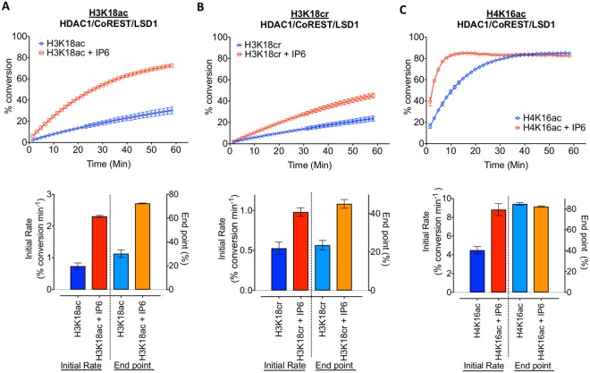
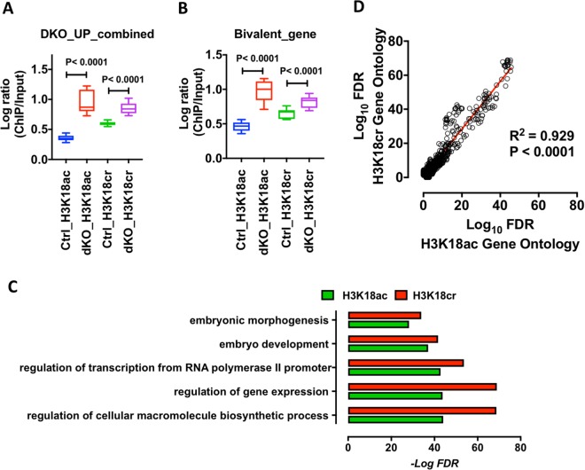
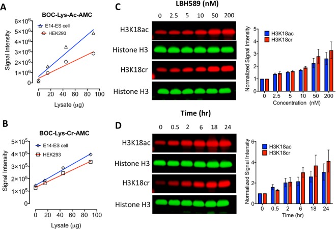
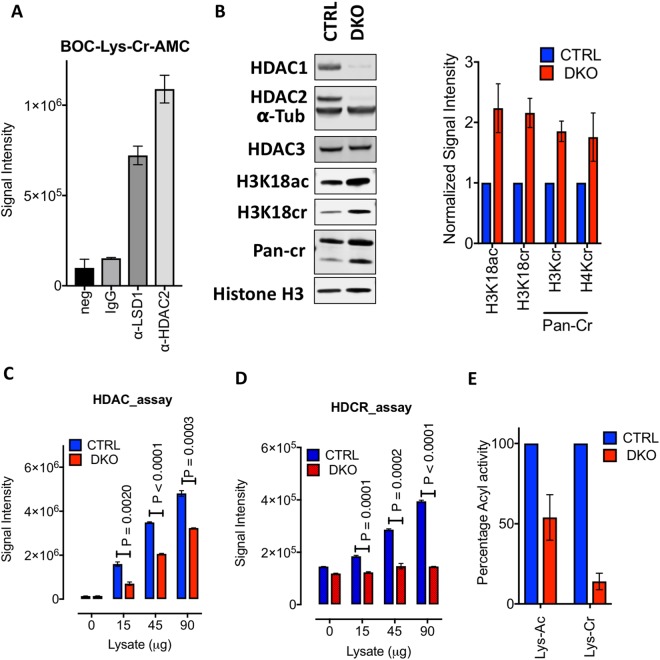
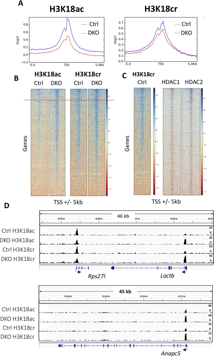
Proteomic analysis of histones has shown that they are subject to a superabundance of acylations, which extend far beyond acetylation, to include: crotonylation, propionylation, butyrylation, malonylation, succinylation, β-hydroxybutyrylation and 2-hydroxyisobutyrylation. To date, much of the functional data has focussed on histone crotonylation which, similar to acetylation, has been associated with positive gene regulation and is added by the acyltransferase, p300. Although Sirtuins 1-3, along with HDAC3, have been shown to possess decrotonylase activity in vitro, there is relatively little known about the regulation of histone crotonylation in vivo. Here we show that Histone Deacetylase 1 and 2 (HDAC1/2), the catalytic core of numerous co-repressor complexes, are important histone decrotonylase enzymes. A ternary complex of HDAC1/CoREST1/LSD1 is able to hydrolyse both histone H3 Lys18-acetyl (H3K18ac) and H3 Lys18-crotonyl (H3K18cr) peptide substrates. Genetic deletion of HDAC1/2 in ES cells increases global levels of histone crotonylation and causes an 85% reduction in total decrotonylase activity. Furthermore, we mapped H3K18cr in cells using ChIP-seq, with and without HDAC1/2, and observed increased levels of crotonylation, which largely overlaps with H3K18ac in the vicinity of transcriptional start sites. Collectively, our data indicate that HDAC1/2 containing complexes are critical regulators of histone crotonylation in vivo.
组蛋白的蛋白质组学分析表明,它们存在大量的酰化修饰,这些修饰远远超出了乙酰化修饰,包括:巴豆酰化、丙酰化、丁酰化、丙二酰化、琥珀酰化、β-羟基丁酰化和 2-羟基异丁酰化。迄今为止,大量的功能数据都集中在组蛋白巴豆酰化上,类似于乙酰化,它与正向基因调控有关,并且是由酰基转移酶 p300 添加的。尽管 Sirtuins 1-3 以及 HDAC3 已被证明在体外具有脱巴豆酰酶活性,但对于组蛋白巴豆酰化的体内调控知之甚少。在这里,我们表明组蛋白去乙酰化酶 1 和 2(HDAC1/2),作为许多共抑制复合物的催化核心,是重要的组蛋白脱巴豆酰酶酶。HDAC1/CoREST1/LSD1 的三元复合物能够水解组蛋白 H3 Lys18-乙酰基(H3K18ac)和 H3 Lys18-巴豆酰基(H3K18cr)肽底物。在 ES 细胞中遗传缺失 HDAC1/2 会增加组蛋白巴豆酰化的整体水平,并导致总脱巴豆酰酶活性降低 85%。此外,我们使用 ChIP-seq 并在有和没有 HDAC1/2 的情况下在细胞中绘制了 H3K18cr 的图谱,观察到巴豆酰化水平升高,这在转录起始位点附近很大程度上与 H3K18ac 重叠。总的来说,我们的数据表明含有 HDAC1/2 的复合物是组蛋白巴豆酰化在体内的关键调节因子。