Faculty of Medicine, Department of Clinical Sciences Lund, Orthopedics, Clinical Epidemiology Unit, Lund University, Lund, Sweden.
Clinical Epidemiology Research & Training Unit, Boston University School of Medicine, Boston, MA, USA.
Eur Radiol. 2019 Apr;29(4):1848-1854. doi: 10.1007/s00330-018-5741-3. Epub 2018 Oct 2.
To determine meniscal extrusion and cartilage coverage on magnetic resonance (MR) images and factors associated with these parameters in knees of middle-aged and elderly persons free from radiographic tibiofemoral osteoarthritis (OA).
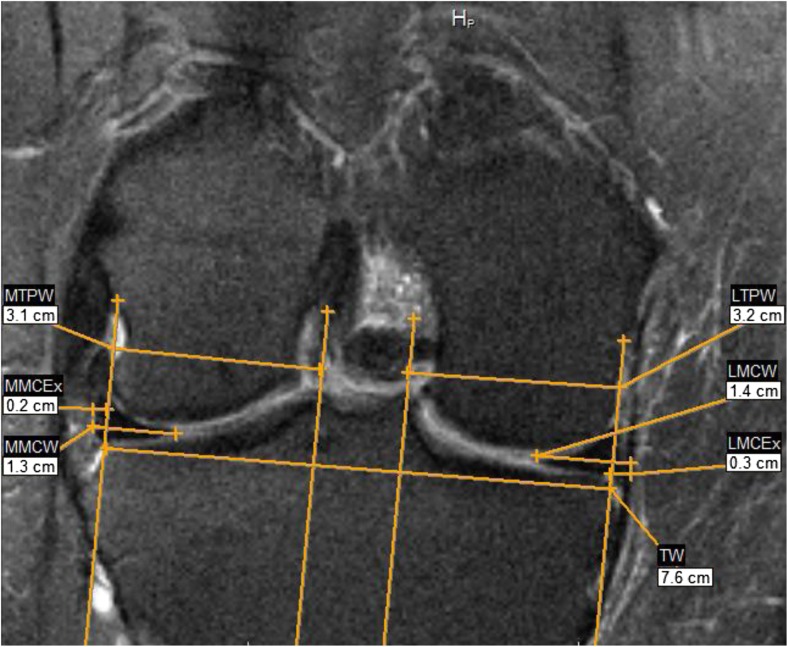
Seven hundred eighteen persons, free of radiographic tibiofemoral OA, aged 50-90 years from Framingham, MA, USA, were included. We measured meniscal extrusion on 1.5 T MRI of both knees to evaluate both medial and lateral meniscal body extrusion and cartilage coverage. We also determined meniscal morphology and structural integrity. The multivariable association with age, body mass index (BMI), and ipsilateral meniscal damage was also evaluated.
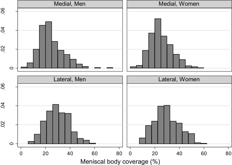
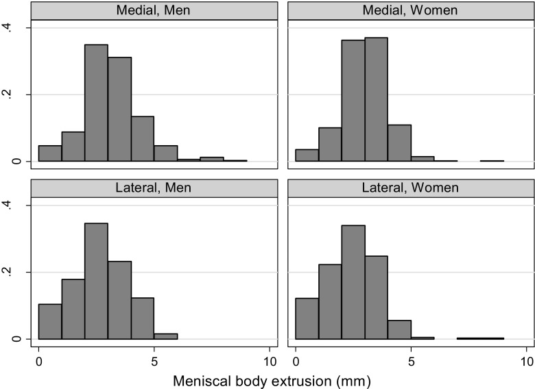
The mean meniscal body extrusion medially was 2.7 mm and laterally 1.8 mm. The tibial cartilage coverage was about 30% of ipsilateral cartilage surface (both compartments). The presence of ipsilateral meniscal damage was associated with more extrusion in only the medial compartment, 1.0 mm in men and 0.6 mm in women, and less cartilage coverage proportion, -5.5% in men and -4.6% in women.
Mean medial meniscal body extrusion in middle-aged or older persons without radiographic tibiofemoral OA approximates the commonly used cutoff (3 mm) to denote pathological extrusion. Medial meniscal damage is a factor associated with medial meniscal body extrusion and less cartilage coverage.
• Medial meniscal extrusion in middle-aged/older persons without OA is around 3 mm. • Lateral meniscal extrusion in middle-aged/older persons without OA is around 2 mm. • Meniscal damage is associated with medial meniscal extrusion and less cartilage coverage.
在磁共振(MR)图像上确定半月板挤出和软骨覆盖,并确定无放射学胫股关节炎(OA)的中老年人膝关节中与这些参数相关的因素。
纳入美国马萨诸塞州弗雷明汉 718 名年龄在 50-90 岁、无放射学胫股 OA 的人群,对其双膝进行 1.5T MRI 检查以评估内侧和外侧半月板体挤出和软骨覆盖情况。我们还确定了半月板形态和结构完整性。还评估了与年龄、体重指数(BMI)和同侧半月板损伤的多变量关联。
内侧半月板体挤出的平均值为 2.7mm,外侧为 1.8mm。胫骨软骨覆盖约为同侧软骨表面的 30%(两个间隔)。同侧半月板损伤的存在仅与内侧间隔的更多挤出有关,男性为 1.0mm,女性为 0.6mm,软骨覆盖比例减少,男性为-5.5%,女性为-4.6%。
无放射学胫股 OA 的中老年人内侧半月板体挤出平均值接近常用的(3mm)病理性挤出截断值。内侧半月板损伤是与内侧半月板体挤出和软骨覆盖减少相关的因素。
• 无 OA 的中老年人的内侧半月板挤出约为 3mm。• 无 OA 的中老年人的外侧半月板挤出约为 2mm。• 半月板损伤与内侧半月板体挤出和软骨覆盖减少有关。