Das Joe M, Abraham Mathew, Nandeesh Bevinahalli N, Nair Suresh N
Department of Neurosurgery, Sree Chitra Tirunal Institute for Medical Sciences and Technology, Thiruvananthapuram, Kerala, India.
Department of Neuropathology, National Institute of Mental Health and Neurosciences, Bengaluru, Karnataka, India.
Asian J Neurosurg. 2018 Jul-Sep;13(3):873-876. doi: 10.4103/ajns.AJNS_350_16.
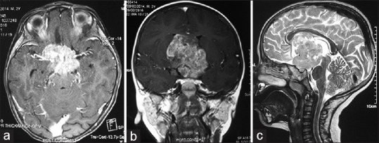
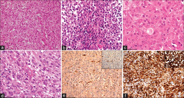
Atypical teratoid rhabdoid tumor (ATRT) is a rare, highly malignant tumor of the central nervous system, commonly affecting children below 3 years of age, with around 300 cases reported in the literature. Suprasellar area is a very rare location for such tumor in the pediatric population, with technical difficulties in complete excision. Third ventricular ATRT is very rare. Here, we report the case of a 2-year-old male child who presented with lethargy and vomiting. He had features of raised intracranial pressure with reduced vision in both eyes. Magnetic resonance imaging of the brain revealed a heterogeneously enhancing lobulated giant lesion in the suprasellar location, occupying the third ventricle and hypothalamus with encasement of both carotids. He underwent pericoronal parasagittal craniotomy, interhemispheric transcallosal interforniceal approach and gross total excision of the lesion. Postoperatively, the child had altered sensorium and diabetes insipidus, both of which recovered over a span of 10 days. Histopathological examination of the specimen was consistent with the diagnosis of World Health Organization Grade IV ATRT. In spite of all our efforts, he succumbed to his illness 5 months postoperatively.
非典型畸胎样横纹肌样瘤(ATRT)是一种罕见的、高度恶性的中枢神经系统肿瘤,常见于3岁以下儿童,文献报道约300例。在儿童人群中,鞍上区域是此类肿瘤非常罕见的发病部位,完整切除存在技术困难。第三脑室ATRT极为罕见。在此,我们报告一例2岁男童,表现为嗜睡和呕吐。他有颅内压升高及双眼视力下降的症状。脑部磁共振成像显示鞍上部位有一个不均匀强化的分叶状巨大病变,占据第三脑室和下丘脑,并包绕双侧颈动脉。他接受了冠状缝旁矢状开颅术、经胼胝体间穹窿间入路,病变被大体全切。术后,患儿出现意识改变和尿崩症,两者均在10天内恢复。标本的组织病理学检查与世界卫生组织IV级ATRT的诊断相符。尽管我们尽了全力,但他在术后5个月因病死亡。