Luo Ji-Zhuang, Zhan Cheng, Ni Xiang, Shi Yu, Wang Qun
Department of Thoracic Surgery, Zhongshan Hospital, Fudan University, Shanghai, 200032, People's Republic of China.
Department of Pathology, Zhongshan Hospital, Fudan University, Shanghai, 200032, People's Republic of China.
J Cardiothorac Surg. 2018 Oct 1;13(1):99. doi: 10.1186/s13019-018-0787-5.
Primary pulmonary meningioma (PPM) is an extremely rare benign tumor. Previous reports indicated that CT features of PPM are single, solid, well-demarcated, homogeneous mass. In this study, we report a case of PPM with atypical CT features.
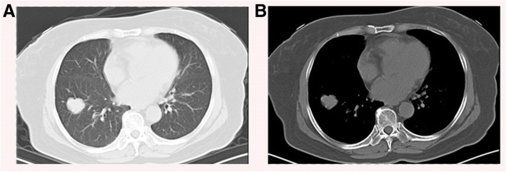
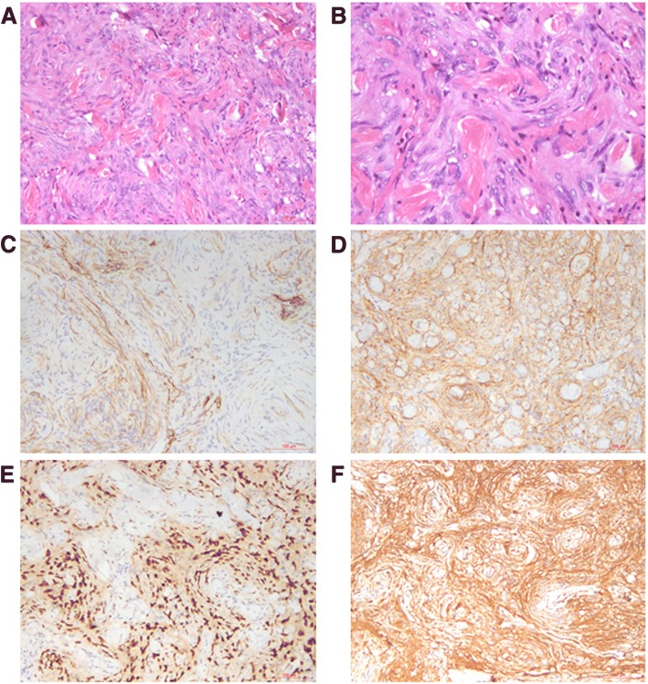
A 65-year-old female presents to clinic with 1-week acute upper respiratory tract infection. Her chest CT scan revealed a 25-29 mm, round-like, heterogeneous lobulated solitary pulmonary nodule in the right lower lobe. Based on the microscopic features and a wide range of immunohistochemical examinations including vimentine, progesterone receptor (PR), CD34 and S100, the mass was diagnosed as PPM after surgery.
PPM is a rare disease, CT features of PPM could be heterogeneous and lobulated. Expression of vimentine, PR, CD34 and S100 helps to diagnosis of PPM.
原发性肺脑膜瘤(PPM)是一种极其罕见的良性肿瘤。既往报道表明,PPM的CT特征为单发、实性、边界清晰、密度均匀的肿块。在本研究中,我们报告1例具有非典型CT特征的PPM病例。
一名65岁女性因1周的急性上呼吸道感染就诊。她的胸部CT扫描显示右肺下叶有一个25 - 29毫米、类圆形、不均匀分叶状的孤立性肺结节。根据显微镜下特征以及包括波形蛋白、孕激素受体(PR)、CD34和S100在内的广泛免疫组化检查结果,该肿块在手术后被诊断为PPM。
PPM是一种罕见疾病,其CT特征可能是不均匀和分叶状的。波形蛋白、PR、CD34和S100的表达有助于PPM的诊断。