Department of Radiation Oncology, Tohoku University Graduate School of Medicine, Sendai, Japan.
Department of Diagnostic Radiology, Tohoku University Graduate School of Medicine, Sendai, Japan.
PLoS One. 2018 Oct 4;13(10):e0204734. doi: 10.1371/journal.pone.0204734. eCollection 2018.
Radiographic severity of radiation-induced lung injury (RILI) has not been well-studied. The goal of this study was to assess the CT appearance pattern and severity of RILI without consideration of the clinical presentation.
A total of 49 patients, 41 with primary lung cancer and 8 with metastatic lung cancer, were treated by 4-fraction stereotactic body radiotherapy (SBRT). RILI after SBRT was separately assessed by two observers. The early and late CT appearance patterns and CT-based severity grading were explored.
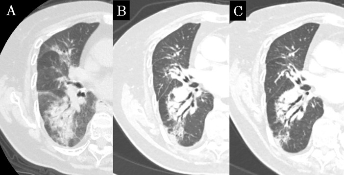
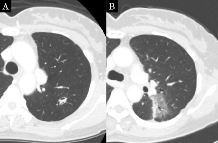
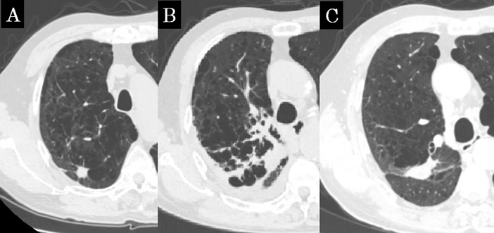
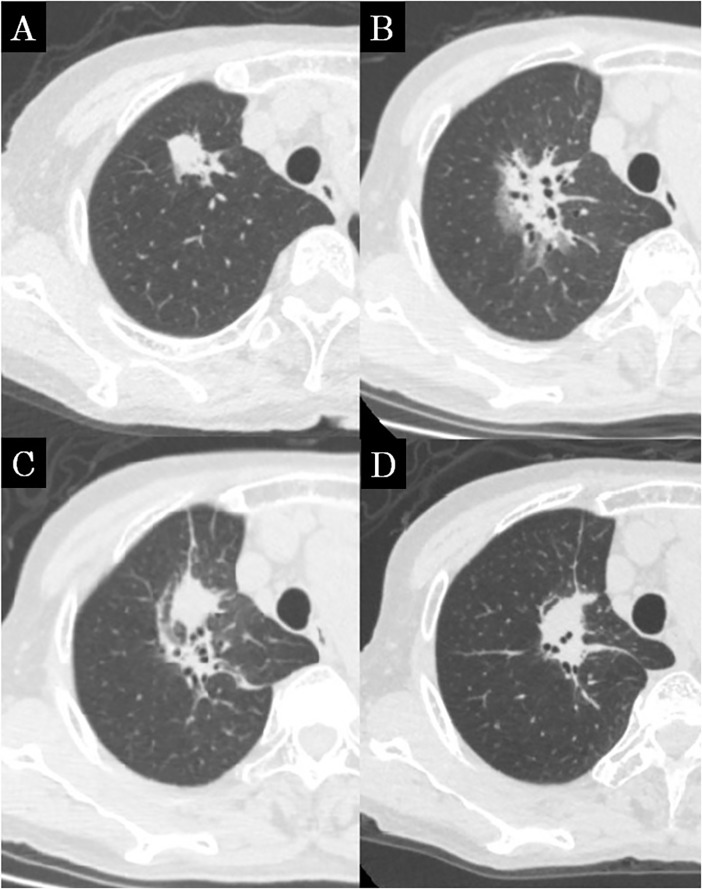
The median follow-up period was 39.0 months. In the early CT findings of observers 1 and 2, there was diffuse consolidation in 15 and 8, diffuse ground glass opacity (GGO) in 0 and 0, patchy consolidation and GGO in 17 and 20, patchy GGO in 3 and 3, and no changes in 10 and 14, respectively (kappa = 0.61). In late CT findings of observer 1 and 2, there were modified conventional pattern in 28 and 24, mass-like pattern in 8 and 11, scar-like pattern in 12 and 12, and no changes in 1 and 2, respectively (kappa = 0.63). In the results of the CT-based grading by observers 1 and 2, there were grade 0 in 1 and 2, grade 1 in 10 and 14, grade 2 in 31 and 29, grade 3 in 7 and 4, and none of grade 4 or more, respectively (kappa = 0.66). According to multivariate analyses (MVA), the significant predicting factors of grade 2 or more CT-based RILI were age (p = 0.01), oxygen dependence (p = 0.03) and interstitial shadow (p = 0.03).
The agreement of the CT appearance and CT-based grading between two observers was good. These indicators may be able to provide us with more objective information and a better understanding of RILI.
放射性肺损伤(RILI)的放射学严重程度尚未得到充分研究。本研究的目的是评估 RILI 的 CT 表现模式和严重程度,而不考虑临床表现。
共纳入 49 例患者,其中 41 例为原发性肺癌,8 例为转移性肺癌,均接受 4 分次立体定向体部放疗(SBRT)。两名观察者分别评估 SBRT 后的 RILI。探讨了早期和晚期 CT 表现模式以及基于 CT 的严重程度分级。
中位随访时间为 39.0 个月。在观察者 1 和 2 的早期 CT 表现中,15 例和 8 例为弥漫性实变,0 例和 0 例为弥漫性磨玻璃密度(GGO),17 例和 20 例为斑片状实变和 GGO,3 例和 3 例为斑片状 GGO,10 例和 14 例无变化(kappa = 0.61)。观察者 1 和 2 的晚期 CT 表现中,28 例和 24 例为改良常规模式,8 例和 11 例为肿块样模式,12 例和 12 例为瘢痕样模式,1 例和 2 例无变化(kappa = 0.63)。观察者 1 和 2 的基于 CT 的分级结果中,1 例和 2 例为 0 级,10 例和 14 例为 1 级,31 例和 29 例为 2 级,7 例和 4 例为 3 级,均无 4 级或更高级别(kappa = 0.66)。多变量分析(MVA)显示,2 级或更高级别的 CT 基于 RILI 的显著预测因素为年龄(p = 0.01)、氧依赖(p = 0.03)和间质影(p = 0.03)。
两名观察者的 CT 表现和基于 CT 的分级一致性良好。这些指标可能为我们提供更客观的信息,并更好地了解 RILI。