Hirsch Gabriella V, Bauer Corinna M, Merabet Lotfi B
The Laboratory for Visual Neuroplasticity, Department of Ophthalmology, Massachusetts Eye and Ear Infirmary, Harvard Medical School. Boston, MA, USA.
Ann Neurosci Psychol. 2015;2. Epub 2015 Aug 13.
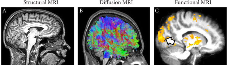
Advances in neuroimaging technology have been instrumental in uncovering the dramatic neurological changes that result from blindness, as well as revealing the inner workings of the human brain. Specifically, modern imaging techniques enable us to examine how the brain adapts and "re-wires" itself as a result of changes in behavior, the environment, injury, or disease; a process referred to as neuroplasticity. Following an overview of commonly employed neuroimaging techniques, we discuss structural and functional neuroplastic brain changes associated with profound visual deprivation. In particular, we highlight how associated structural changes often occur within areas that process intact senses (such as hearing, touch, and smell) while functional changes tend to implicate areas of the brain normally ascribed to the processing of visual information. Evidence will primarily focus on profound blindness due to ocular cause, but related work in cerebral/cortical visual impairment (CVI) will also be discussed. The potential importance of these findings within the context of education and rehabilitation is proposed.
神经成像技术的进步有助于揭示因失明而导致的显著神经变化,同时也能揭示人类大脑的内部运作机制。具体而言,现代成像技术使我们能够研究大脑如何因行为、环境、损伤或疾病的变化而进行自我适应和“重新布线”,这一过程被称为神经可塑性。在概述常用的神经成像技术之后,我们将讨论与严重视觉剥夺相关的神经可塑性大脑的结构和功能变化。特别是,我们将重点强调相关结构变化通常发生在处理完整感官(如听觉、触觉和嗅觉)的区域内,而功能变化往往涉及通常归因于视觉信息处理的大脑区域。证据将主要集中于因眼部原因导致的严重失明,但也会讨论在脑/皮质视觉障碍(CVI)方面的相关研究。本文还提出了这些发现在教育和康复背景下的潜在重要性。