Chen Tzu-Pai, Yi Wan-Ling, Liu Chiang-Shin, Lin Yu-Hsuan
Medical College of National Cheng Kung University.
Department of Pathology.
Medicine (Baltimore). 2018 Oct;97(40):e12615. doi: 10.1097/MD.0000000000012615.
Xanthogranulomatous inflammation (XGI) is a rare inflammatory process, which mostly affects the kidney and gallbladder. It usually simulates an aggressive neoplastic process. Occurrences in the neck are extremely rare and would usually be associated with a preexisting cyst or glandular tissues.
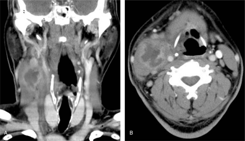
A 49-year-old diabetic patient presented with a right painful neck mass for a week. The pretreatment computed tomography (CT) imaging with contrast demonstrated a huge ill-defined heterogeneous-enhanced lesion abutting surrounding musculatures and great vessels. Both fine needle aspiration (FNA) and ultrasound-guided core biopsy of the neck mass showed inflammatory cells only.
Histologic evaluation found granulation tissue with histiocytes and occasional Touton giant cells confirming the diagnosis of xanthogranuloma.
Open excisional biopsy demonstrated a yellowish mass-like lesion with abscess inside.
The patient recovered from the disease without posttreatment comorbidities.
This case highlights the need for physicians to maintain awareness of this clinical entity and delayed- or overtreatment should be avoided in these patients due to preoperative ambiguous diagnosis.
黄色肉芽肿性炎症(XGI)是一种罕见的炎症过程,主要累及肾脏和胆囊。它通常类似侵袭性肿瘤过程。颈部发生极为罕见,通常与先前存在的囊肿或腺组织有关。
一名49岁糖尿病患者出现右侧颈部疼痛性肿块一周。术前增强计算机断层扫描(CT)成像显示一个巨大的边界不清的不均匀强化病变,毗邻周围肌肉组织和大血管。颈部肿块的细针穿刺抽吸(FNA)和超声引导下的粗针活检均仅显示炎性细胞。
组织学评估发现有肉芽组织,伴有组织细胞和偶见的杜顿巨细胞,确诊为黄色肉芽肿。
开放性切除活检显示为一个黄色的肿块样病变,内部有脓肿。
患者康复,无治疗后合并症。
该病例强调医生需要对这种临床实体保持警惕,由于术前诊断不明确,应避免对这些患者进行延迟治疗或过度治疗。