• 文献检索
  • 文档翻译
  • 深度研究
  • 学术资讯
  • Suppr Zotero 插件Zotero 插件
  • 邀请有礼
  • 套餐&价格
  • 历史记录
应用&插件
Suppr Zotero 插件Zotero 插件浏览器插件Mac 客户端Windows 客户端微信小程序
定价
高级版会员购买积分包购买API积分包
服务
文献检索文档翻译深度研究API 文档MCP 服务
关于我们
关于 Suppr公司介绍联系我们用户协议隐私条款
关注我们

Suppr 超能文献

核心技术专利:CN118964589B侵权必究
粤ICP备2023148730 号-1Suppr @ 2026

文献检索

告别复杂PubMed语法,用中文像聊天一样搜索,搜遍4000万医学文献。AI智能推荐,让科研检索更轻松。

立即免费搜索

文件翻译

保留排版,准确专业,支持PDF/Word/PPT等文件格式,支持 12+语言互译。

免费翻译文档

深度研究

AI帮你快速写综述,25分钟生成高质量综述,智能提取关键信息,辅助科研写作。

立即免费体验

基于 HoloLens 的高分辨率心肌瘢痕的三维全息可视化。

Three-dimensional holographic visualization of high-resolution myocardial scar on HoloLens.

机构信息

Department of Medicine (Cardiovascular Division), Beth Israel Deaconess Medical Center and Harvard Medical School, Boston, MA, United States of America.

Department of Computer Science, Technical University of Munich, Munich, Germany.

出版信息

PLoS One. 2018 Oct 8;13(10):e0205188. doi: 10.1371/journal.pone.0205188. eCollection 2018.

DOI:10.1371/journal.pone.0205188
PMID:30296291
原文链接:https://pmc.ncbi.nlm.nih.gov/articles/PMC6175509/
Abstract

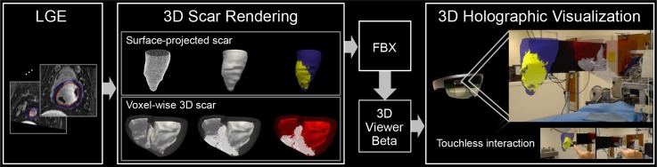
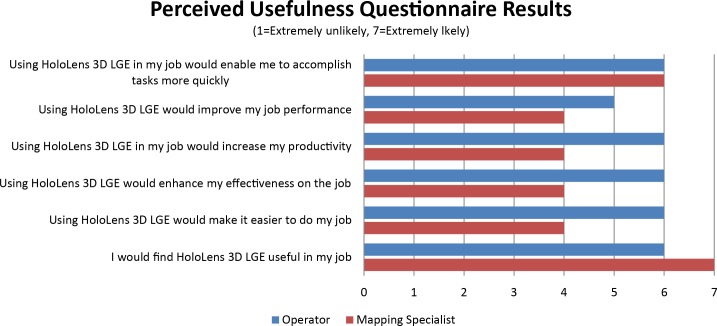
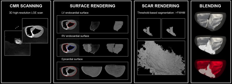
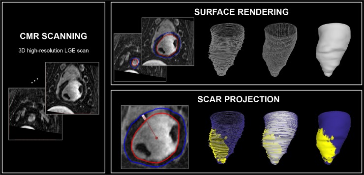
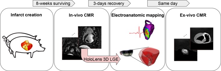
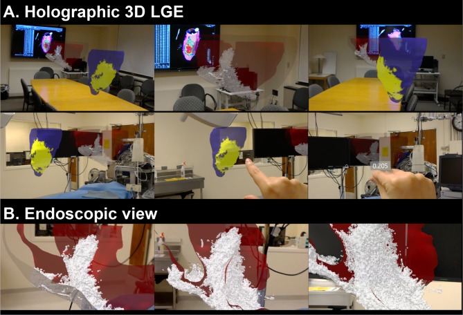
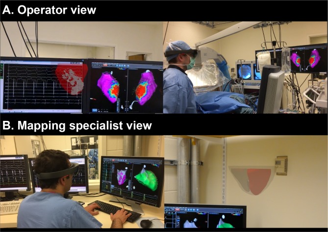
Visualization of the complex 3D architecture of myocardial scar could improve guidance of radio-frequency ablation in the treatment of ventricular tachycardia (VT). In this study, we sought to develop a framework for 3D holographic visualization of myocardial scar, imaged using late gadolinium enhancement (LGE), on the augmented reality HoloLens. 3D holographic LGE model was built using the high-resolution 3D LGE image. Smooth endo/epicardial surface meshes were generated using Poisson surface reconstruction. For voxel-wise 3D scar model, every scarred voxel was rendered into a cube which carries the actual resolution of the LGE sequence. For surface scar model, scar information was projected on the endocardial surface mesh. Rendered layers were blended with different transparency and color, and visualized on HoloLens. A pilot animal study was performed where 3D holographic visualization of the scar was performed in 5 swines who underwent controlled infarction and electroanatomic mapping to identify VT substrate. 3D holographic visualization enabled assessment of the complex 3D scar architecture with touchless interaction in a sterile environment. Endoscopic view allowed visualization of scar from the ventricular chambers. Upon completion of the animal study, operator and mapping specialist independently completed the perceived usefulness questionnaire in the six-item usefulness scale. Operator and mapping specialist found it useful (usefulness rating: operator, 5.8; mapping specialist, 5.5; 1-7 scale) to have scar information during the intervention. HoloLens 3D LGE provides a true 3D perception of the complex scar architecture with immersive experience to visualize scar in an interactive and interpretable 3D approach, which may facilitate MR-guided VT ablation.

摘要

心肌瘢痕的复杂 3D 结构可视化可以改善射频消融治疗室性心动过速 (VT) 的指导。在这项研究中,我们试图开发一种基于增强现实 HoloLens 的心肌瘢痕 3D 全息可视化框架,使用晚期钆增强 (LGE) 进行成像。使用高分辨率 3D LGE 图像构建 3D 全息 LGE 模型。使用泊松曲面重建生成光滑的心内膜/心外膜表面网格。对于体素化 3D 瘢痕模型,将每个瘢痕化体素渲染成一个带有 LGE 序列实际分辨率的立方体。对于表面瘢痕模型,将瘢痕信息投影到心内膜表面网格上。渲染层以不同的透明度和颜色混合,并在 HoloLens 上可视化。进行了一项初步动物研究,在 5 头接受控制性梗死和电解剖标测以识别 VT 底物的猪中进行了瘢痕的 3D 全息可视化。3D 全息可视化允许在无菌环境中进行非接触式交互评估复杂的 3D 瘢痕结构。内窥镜视图允许从心室腔观察瘢痕。动物研究完成后,操作人员和绘图专家分别在六项有用性量表中完成了对感知有用性的问卷调查。操作人员和绘图专家发现,在干预过程中获取瘢痕信息非常有用(有用性评分:操作人员 5.8;绘图专家 5.5;1-7 量表)。HoloLens 3D LGE 以沉浸式体验提供了对复杂瘢痕结构的真实 3D 感知,以交互式和可解释的 3D 方式可视化瘢痕,这可能有助于磁共振引导的 VT 消融。

https://cdn.ncbi.nlm.nih.gov/pmc/blobs/bea3/6175509/2b675ed05439/pone.0205188.g007.jpg
https://cdn.ncbi.nlm.nih.gov/pmc/blobs/bea3/6175509/5dec2ee9055d/pone.0205188.g001.jpg
https://cdn.ncbi.nlm.nih.gov/pmc/blobs/bea3/6175509/2adff8547359/pone.0205188.g002.jpg
https://cdn.ncbi.nlm.nih.gov/pmc/blobs/bea3/6175509/45967e1c179a/pone.0205188.g003.jpg
https://cdn.ncbi.nlm.nih.gov/pmc/blobs/bea3/6175509/e49927ae0ae5/pone.0205188.g004.jpg
https://cdn.ncbi.nlm.nih.gov/pmc/blobs/bea3/6175509/86d96c6686c9/pone.0205188.g005.jpg
https://cdn.ncbi.nlm.nih.gov/pmc/blobs/bea3/6175509/3b61123560bf/pone.0205188.g006.jpg
https://cdn.ncbi.nlm.nih.gov/pmc/blobs/bea3/6175509/2b675ed05439/pone.0205188.g007.jpg

相似文献

1
Three-dimensional holographic visualization of high-resolution myocardial scar on HoloLens.基于 HoloLens 的高分辨率心肌瘢痕的三维全息可视化。
PLoS One. 2018 Oct 8;13(10):e0205188. doi: 10.1371/journal.pone.0205188. eCollection 2018.
2
Integration of merged delayed-enhanced magnetic resonance imaging and multidetector computed tomography for the guidance of ventricular tachycardia ablation: a pilot study.融合延迟强化磁共振成像与多排 CT 用于指导室性心动过速消融术:一项初步研究。
J Cardiovasc Electrophysiol. 2013 Apr;24(4):419-26. doi: 10.1111/jce.12052. Epub 2012 Dec 17.
3
MRI-Guided ventricular tachycardia ablation: integration of late gadolinium-enhanced 3D scar in patients with implantable cardioverter-defibrillators.MRI 引导下心室性心动过速消融术:在植入式心脏复律除颤器患者中整合晚期钆增强 3D 瘢痕。
Circ Arrhythm Electrophysiol. 2011 Apr;4(2):172-84. doi: 10.1161/CIRCEP.110.958744. Epub 2011 Jan 26.
4
Regional Strain by Cardiac Magnetic Resonance Imaging Improves Detection of Right Ventricular Scar Compared With Late Gadolinium Enhancement on a Multimodality Scar Evaluation in Patients With Arrhythmogenic Right Ventricular Cardiomyopathy.心脏磁共振成像的区域性应变与多模态瘢痕评估中的晚期钆增强相比,可提高心律失常性右心室心肌病患者右心室瘢痕的检出率。
Circ Cardiovasc Imaging. 2018 Sep;11(9):e007546. doi: 10.1161/CIRCIMAGING.118.007546.
5
Three-dimensional architecture of scar and conducting channels based on high resolution ce-CMR: insights for ventricular tachycardia ablation.基于高分辨率 ce-CMR 的瘢痕和传导通道的三维结构:对室性心动过速消融的启示。
Circ Arrhythm Electrophysiol. 2013 Jun;6(3):528-37. doi: 10.1161/CIRCEP.113.000264. Epub 2013 May 17.
6
3D delayed-enhanced magnetic resonance sequences improve conducting channel delineation prior to ventricular tachycardia ablation.三维延迟增强磁共振序列可改善室性心动过速消融术前传导通道的描绘。
Europace. 2015 Jun;17(6):938-45. doi: 10.1093/europace/euu310. Epub 2015 Jan 23.
7
Device artifact reduction for magnetic resonance imaging of patients with implantable cardioverter-defibrillators and ventricular tachycardia: late gadolinium enhancement correlation with electroanatomic mapping.植入式心脏复律除颤器和室性心动过速患者磁共振成像的器械伪影减少:晚期钆增强与电解剖标测的相关性。
Heart Rhythm. 2014 Feb;11(2):289-98. doi: 10.1016/j.hrthm.2013.10.032. Epub 2013 Oct 16.
8
Improved multimodality data fusion of late gadolinium enhancement MRI to left ventricular voltage maps in ventricular tachycardia ablation.提高室性心动过速消融术中心肌延迟强化 MRI 与左心室电压图的多模态数据融合
IEEE Trans Biomed Eng. 2013 May;60(5):1308-17. doi: 10.1109/TBME.2012.2233738. Epub 2012 Dec 12.
9
Feasibility of image-based simulation to estimate ablation target in human ventricular arrhythmia.基于影像的模拟技术估计人类室性心律失常消融靶点的可行性。
Heart Rhythm. 2013 Aug;10(8):1109-16. doi: 10.1016/j.hrthm.2013.04.015. Epub 2013 Apr 19.
10
A swine model of infarct-related reentrant ventricular tachycardia: Electroanatomic, magnetic resonance, and histopathological characterization.梗死相关折返性室性心动过速的猪模型:电解剖、磁共振及组织病理学特征
Heart Rhythm. 2016 Jan;13(1):262-73. doi: 10.1016/j.hrthm.2015.07.030. Epub 2015 Jul 28.

引用本文的文献

1
Mixed Reality (Holography)-Guided Minimally Invasive Cardiac Surgery-A Novel Comparative Feasibility Study.混合现实(全息技术)引导下的微创心脏手术——一项新型比较可行性研究
J Cardiovasc Dev Dis. 2025 Jan 27;12(2):49. doi: 10.3390/jcdd12020049.
2
First Experience With Augmented Reality Guidance for Cerebral Embolic Protection During TAVR.经导管主动脉瓣置换术(TAVR)期间使用增强现实引导进行脑栓塞保护的首次经验。
JACC Adv. 2024 Feb 7;3(3):100839. doi: 10.1016/j.jacadv.2024.100839. eCollection 2024 Mar.
3
Visions for digital integrated cardiovascular care: HRS Digital Health Committee perspectives.

本文引用的文献

1
Cardiac MR Characterization of left ventricular remodeling in a swine model of infarct followed by reperfusion.心肌梗死后再灌注猪模型中左心室重构的心脏磁共振特征分析
J Magn Reson Imaging. 2018 Mar 9. doi: 10.1002/jmri.26005.
2
Standard Ablation Versus Magnetic Resonance Imaging-Guided Ablation in the Treatment of Ventricular Tachycardia.标准消融与磁共振成像引导下消融治疗室性心动过速的比较
Circ Arrhythm Electrophysiol. 2018 Jan;11(1):e005973. doi: 10.1161/CIRCEP.117.005973.
3
Towards X-ray free endovascular interventions - using HoloLens for on-line holographic visualisation.
数字集成心血管护理愿景:心脏节律学会数字健康委员会观点
Cardiovasc Digit Health J. 2024 Feb 24;5(2):37-49. doi: 10.1016/j.cvdhj.2024.02.003. eCollection 2024 Apr.
4
Use of a mixed reality system for navigational mapping during cardiac electrophysiological testing does not prolong case duration: A subanalysis from the Cardiac Augmented REality study.在心脏电生理检查期间使用混合现实系统进行导航映射不会延长病例时长:来自心脏增强现实研究的亚分析
Cardiovasc Digit Health J. 2023 Jun 14;4(4):111-117. doi: 10.1016/j.cvdhj.2023.06.003. eCollection 2023 Aug.
5
A System for Real-Time, Online Mixed-Reality Visualization of Cardiac Magnetic Resonance Images.一种用于心脏磁共振图像实时在线混合现实可视化的系统。
J Imaging. 2021 Dec 14;7(12):274. doi: 10.3390/jimaging7120274.
6
A Virtual Reality System for Improved Image-Based Planning of Complex Cardiac Procedures.一种用于改进复杂心脏手术基于图像规划的虚拟现实系统。
J Imaging. 2021 Aug 19;7(8):151. doi: 10.3390/jimaging7080151.
7
Measurement and 3D Visualization of the Human Internal Heat Field by Means of Microwave Radiometry.利用微波辐射计测量和三维可视化人体内部热场。
Sensors (Basel). 2021 Jun 10;21(12):4005. doi: 10.3390/s21124005.
8
Digital health innovation in cardiology.心脏病学中的数字健康创新。
Cardiovasc Digit Health J. 2020 Jul-Aug;1(1):6-8. doi: 10.1016/j.cvdhj.2020.07.003. Epub 2020 Aug 28.
9
3D visualization and 3D printing in abnormal gastrointestinal system manifestations of situs ambiguus.内脏反位异常胃肠道系统表现中的三维可视化与三维打印
Quant Imaging Med Surg. 2020 Sep;10(9):1877-1883. doi: 10.21037/qims-20-661.
10
Experimental Setup Employed in the Operating Room Based on Virtual and Mixed Reality: Analysis of Pros and Cons in Open Abdomen Surgery.基于虚拟现实和混合现实的手术室实验设置:开放性腹部手术中的优缺点分析。
J Healthc Eng. 2020 Aug 6;2020:8851964. doi: 10.1155/2020/8851964. eCollection 2020.
迈向无X射线的血管内介入治疗——利用HoloLens进行在线全息可视化
Healthc Technol Lett. 2017 Sep 14;4(5):184-187. doi: 10.1049/htl.2017.0061. eCollection 2017 Oct.
4
Cardiac magnetic resonance-aided scar dechanneling: Influence on acute and long-term outcomes.心脏磁共振辅助瘢痕去通道化:对急性和长期结局的影响。
Heart Rhythm. 2017 Aug;14(8):1121-1128. doi: 10.1016/j.hrthm.2017.05.018.
5
Clinical performance of high-resolution late gadolinium enhancement imaging with compressed sensing.应用压缩感知技术的高分辨率晚期钆增强成像的临床性能。
J Magn Reson Imaging. 2017 Dec;46(6):1829-1838. doi: 10.1002/jmri.25695. Epub 2017 Mar 16.
6
Touchless interaction with software in interventional radiology and surgery: a systematic literature review.介入放射学与手术中软件的非接触式交互:一项系统的文献综述
Int J Comput Assist Radiol Surg. 2017 Feb;12(2):291-305. doi: 10.1007/s11548-016-1480-6. Epub 2016 Sep 19.
7
Computer-generated real-time digital holography: first time use in clinical medical imaging.计算机生成的实时数字全息术:首次应用于临床医学成像。
Eur Heart J Cardiovasc Imaging. 2016 Aug;17(8):845-9. doi: 10.1093/ehjci/jew087. Epub 2016 Jun 9.
8
Evaluation of the effectiveness of 3D vascular stereoscopic models in anatomy instruction for first year medical students.3D血管立体模型在一年级医学生解剖学教学中的有效性评估。
Anat Sci Educ. 2017 Jan;10(1):34-45. doi: 10.1002/ase.1626. Epub 2016 Jun 6.
9
Holography for imaging in structural heart disease.用于结构性心脏病成像的全息术。
EuroIntervention. 2016 May 17;12 Suppl X:X81-X84. doi: 10.4244/EIJV12SXA15.
10
Augmented reality in neurosurgery: a systematic review.神经外科中的增强现实:一项系统综述。
Neurosurg Rev. 2017 Oct;40(4):537-548. doi: 10.1007/s10143-016-0732-9. Epub 2016 May 7.