Bast Björn-Ole, Rickert Uta, Schneppenheim Janna, Cossais François, Wilms Henrik, Arnold Philipp, Lucius Ralph
Anatomical Institute, Christian-Albrechts-University of Kiel, Otto-Hahn Platz 8, 24118 Kiel, Germany.
Department of Neurology, Texas Tech University Health Science Center, 3601 4th Street, 79430 Lubbock, TX, USA.
Heliyon. 2018 Oct 3;4(10):e00826. doi: 10.1016/j.heliyon.2018.e00826. eCollection 2018 Oct.
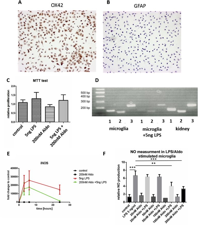
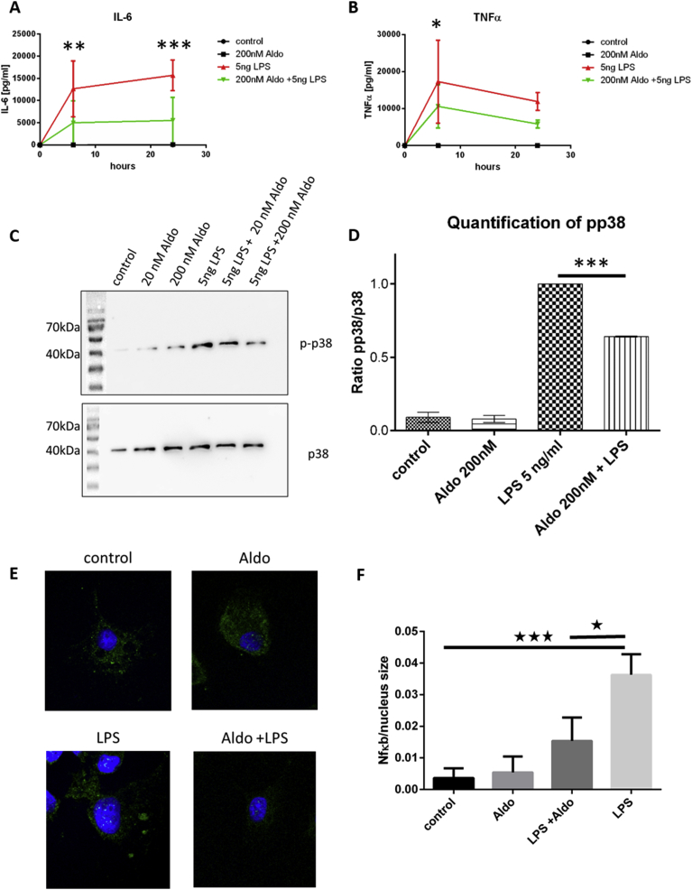
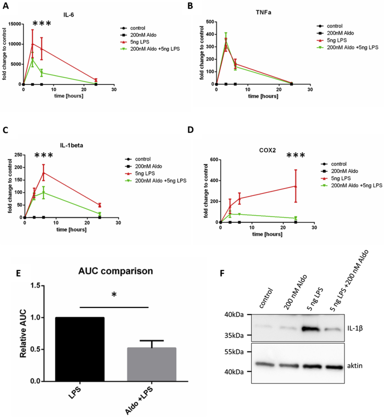
Over the last years, studies on microglia cell function in chronic neuro-inflammation and neuronal necrosis pointed towards an eminent role of these cells in Multiple Sclerosis, Parkinson's and Alzheimer's Disease. It was found, that microglia cell activity can be stimulated towards a pro- or an anti-inflammatory profile, depending on the stimulating signals. Therefore, investigation of receptors expressed by microglia cells and ligands influencing their activation state is of eminent interest. A receptor found to be expressed by microglia cells is the mineralocorticoid receptor. One of its ligands is Aldosterone, a naturally produced steroid hormone of the adrenal cortex, which mainly induces homeostatic and renal effects. We evaluated if the addition of Aldosterone to LPS stimulated microglia cells changes their inflammatory profile. Therefore, we assessed the levels of nitric oxide (NO), iNOS, IL-6, IL-1β, TNF-α and COX-2 in untreated, LPS-treated and LPS/Aldosterone-treated microglia cells. Furthermore we analyzed p38-MAP-Kinase and NFκB signaling within these cells. Our results indicate that the co-stimulation with Aldosterone leads to a decrease of the LPS-induced pro-inflammatory effect and thus renders Aldosterone an anti-inflammatory agent in our model system.
在过去几年中,关于小胶质细胞在慢性神经炎症和神经元坏死中的功能研究表明,这些细胞在多发性硬化症、帕金森病和阿尔茨海默病中起着重要作用。研究发现,根据刺激信号的不同,小胶质细胞的活性可以被刺激为促炎或抗炎状态。因此,研究小胶质细胞表达的受体以及影响其激活状态的配体具有重要意义。已发现小胶质细胞表达的一种受体是盐皮质激素受体。其配体之一是醛固酮,它是肾上腺皮质自然产生的一种类固醇激素,主要诱导体内平衡和肾脏效应。我们评估了向脂多糖(LPS)刺激的小胶质细胞中添加醛固酮是否会改变其炎症状态。因此,我们检测了未处理、LPS处理和LPS/醛固酮处理的小胶质细胞中一氧化氮(NO)、诱导型一氧化氮合酶(iNOS)、白细胞介素-6(IL-6)、白细胞介素-1β(IL-1β)、肿瘤坏死因子-α(TNF-α)和环氧化酶-2(COX-2)的水平。此外,我们分析了这些细胞内的p38丝裂原活化蛋白激酶(p38-MAP-Kinase)和核因子κB(NFκB)信号通路。我们的结果表明,醛固酮的共同刺激导致LPS诱导的促炎作用减弱,因此在我们的模型系统中醛固酮是一种抗炎剂。