• 文献检索
  • 文档翻译
  • 深度研究
  • 学术资讯
  • Suppr Zotero 插件Zotero 插件
  • 邀请有礼
  • 套餐&价格
  • 历史记录
应用&插件
Suppr Zotero 插件Zotero 插件浏览器插件Mac 客户端Windows 客户端微信小程序
定价
高级版会员购买积分包购买API积分包
服务
文献检索文档翻译深度研究API 文档MCP 服务
关于我们
关于 Suppr公司介绍联系我们用户协议隐私条款
关注我们

Suppr 超能文献

核心技术专利:CN118964589B侵权必究
粤ICP备2023148730 号-1Suppr @ 2026

文献检索

告别复杂PubMed语法,用中文像聊天一样搜索,搜遍4000万医学文献。AI智能推荐,让科研检索更轻松。

立即免费搜索

文件翻译

保留排版,准确专业,支持PDF/Word/PPT等文件格式,支持 12+语言互译。

免费翻译文档

深度研究

AI帮你快速写综述,25分钟生成高质量综述,智能提取关键信息,辅助科研写作。

立即免费体验

采用脉搏轮廓法测量被动抬腿试验中每搏量的变化。

Using the Pulse Contour Method to Measure the Changes in Stroke Volume during a Passive Leg Raising Test.

机构信息

Institute of Medicine, School of Medicine, Chung-Shan Medical University; Department of Internal Medicine, Chung-Shan Medical University Hospital, Taichung 402, Taiwan.

Department of Computer Science and Information Engineering, Chaoyang University of Technology, Taichung 413, Taiwan.

出版信息

Sensors (Basel). 2018 Oct 12;18(10):3420. doi: 10.3390/s18103420.

DOI:10.3390/s18103420
PMID:30322018
原文链接:https://pmc.ncbi.nlm.nih.gov/articles/PMC6210240/
Abstract

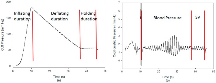
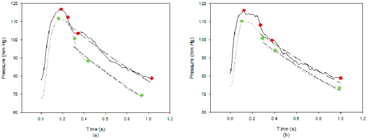
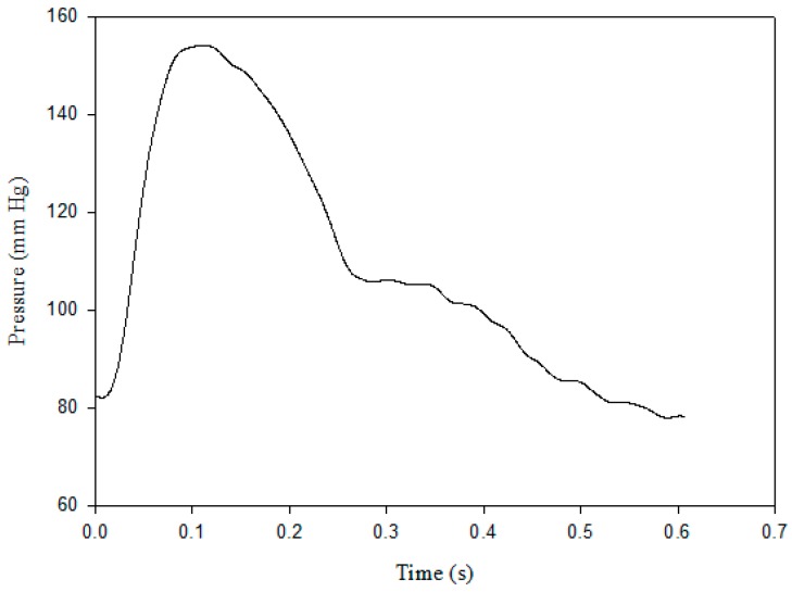
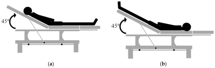
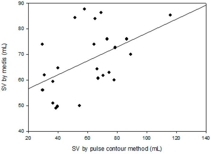
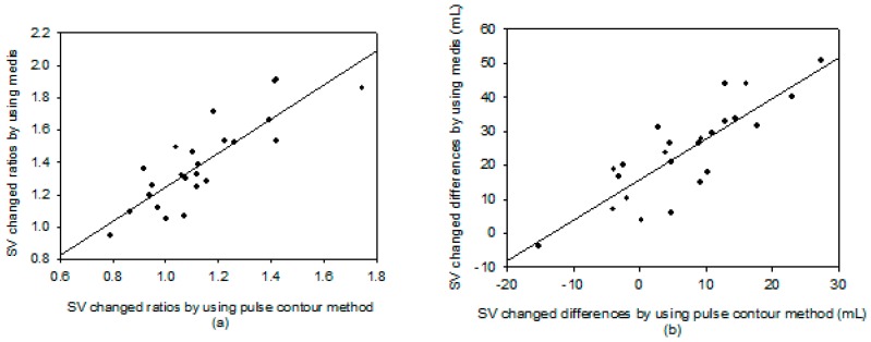
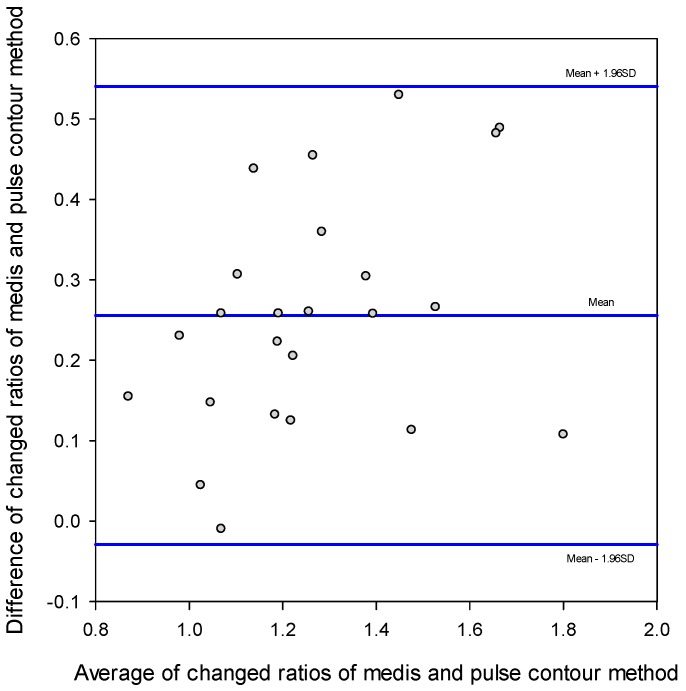
The pulse contour method is often used with the Windkessel model to measure stroke volume. We used a digital pressure and flow sensors to detect the parameters of the Windkessel model from the pulse waveform. The objective of this study was to assess the stability and accuracy of this method by making use of the passive leg raising test. We studied 24 healthy subjects (40 ± 9.3 years), and used the Medis CS 1000, an impedance cardiography, as the comparing reference. The pulse contour method measured the waveform of the brachial artery by using a cuff. The compliance and resistance of the peripheral artery was detected from the cuff characteristics and the blood pressure waveform. Then, according to the method proposed by Romano et al., the stroke volume could be measured. This method was implemented in our designed blood pressure monitor. A passive leg raising test, which could immediately change the preloading of the heart, was done to certify the performance of our method. The pulse contour method and impedance cardiography simultaneously measured the stroke volume. The measurement of the changes in stroke volume using the pulse contour method had a very high correlation with the Medis CS 1000 measurement, the correlation coefficient of the changed ratio and changed differences in stroke volume were ² = 0.712 and ² = 0.709, respectively. It was shown that the stroke volume measured by using the pulse contour method was not accurate enough. But, the changes in the stroke volume could be accurately measured with this pulse contour method. Changes in stroke volume are often used to understand the conditions of cardiac preloading in the clinical field. Moreover, the operation of the pulse contour method is easier than using impedance cardiography and echocardiography. Thus, this method is suitable to use in different healthcare fields.

摘要

脉搏轮廓法常与 Windkessel 模型结合,用于测量心搏量。我们使用数字压力和流量传感器,从脉搏波形中检测 Windkessel 模型的参数。本研究的目的是利用被动抬腿试验评估该方法的稳定性和准确性。我们研究了 24 名健康受试者(40 ± 9.3 岁),并使用 Medis CS 1000 阻抗心动图作为比较参考。脉搏轮廓法使用袖带测量肱动脉的波形。通过袖带特性和血压波形检测外周动脉的顺应性和阻力。然后,根据 Romano 等人提出的方法,可以测量心搏量。该方法在我们设计的血压监测仪中实现。进行被动抬腿试验,可立即改变心脏的前负荷,以验证我们方法的性能。脉搏轮廓法和阻抗心动图同时测量心搏量。脉搏轮廓法测量心搏量变化与 Medis CS 1000 测量具有很高的相关性,心搏量变化比和变化差异的相关系数分别为² = 0.712 和² = 0.709。结果表明,使用脉搏轮廓法测量的心搏量不够准确。但是,使用这种脉搏轮廓法可以准确测量心搏量的变化。心搏量变化常用于了解临床领域中心脏前负荷的情况。此外,脉搏轮廓法的操作比使用阻抗心动图和超声心动图更简单。因此,该方法适用于不同的医疗保健领域。

https://cdn.ncbi.nlm.nih.gov/pmc/blobs/3f3b/6210240/9a75b96d92c3/sensors-18-03420-g007.jpg
https://cdn.ncbi.nlm.nih.gov/pmc/blobs/3f3b/6210240/1b452eec349a/sensors-18-03420-g001.jpg
https://cdn.ncbi.nlm.nih.gov/pmc/blobs/3f3b/6210240/9b13022c7079/sensors-18-03420-g002.jpg
https://cdn.ncbi.nlm.nih.gov/pmc/blobs/3f3b/6210240/9ad5e93f4381/sensors-18-03420-g003.jpg
https://cdn.ncbi.nlm.nih.gov/pmc/blobs/3f3b/6210240/0e382b405658/sensors-18-03420-g004.jpg
https://cdn.ncbi.nlm.nih.gov/pmc/blobs/3f3b/6210240/b6c0935803c2/sensors-18-03420-g005.jpg
https://cdn.ncbi.nlm.nih.gov/pmc/blobs/3f3b/6210240/14b90c31c0db/sensors-18-03420-g006.jpg
https://cdn.ncbi.nlm.nih.gov/pmc/blobs/3f3b/6210240/9a75b96d92c3/sensors-18-03420-g007.jpg

相似文献

1
Using the Pulse Contour Method to Measure the Changes in Stroke Volume during a Passive Leg Raising Test.采用脉搏轮廓法测量被动抬腿试验中每搏量的变化。
Sensors (Basel). 2018 Oct 12;18(10):3420. doi: 10.3390/s18103420.
2
Comparison of pulse contour, aortic Doppler ultrasound and bioelectrical impedance estimates of stroke volume during rapid changes in blood pressure.血压快速变化期间每搏输出量的脉搏轮廓、主动脉多普勒超声及生物电阻抗估计值的比较
Exp Physiol. 2019 Mar;104(3):368-378. doi: 10.1113/EP087240. Epub 2019 Jan 27.
3
Assessment of Stroke Volume From Brachial Blood Pressure Using Arterial Characteristics.利用动脉特征从肱动脉血压评估每搏输出量
IEEE Trans Biomed Eng. 2015 Sep;62(9):2151-7. doi: 10.1109/TBME.2015.2412136. Epub 2015 Mar 19.
4
Improved pressure contour analysis for estimating cardiac stroke volume using pulse wave velocity measurement.利用脉搏波速度测量改进压力轮廓分析以估计心搏量
Biomed Eng Online. 2017 Apr 24;16(1):51. doi: 10.1186/s12938-017-0341-z.
5
Monitoring of cardiac output by pulse contour method.通过脉搏轮廓法监测心输出量。
Acta Anaesthesiol Belg. 1978;29(3):259-70.
6
The effect of variable arterial transducer level on the accuracy of pulse contour waveform-derived measurements in critically ill patients.动脉换能器水平变化对危重症患者脉搏轮廓波形衍生测量准确性的影响。
J Clin Monit Comput. 2016 Oct;30(5):569-75. doi: 10.1007/s10877-015-9756-x. Epub 2015 Aug 23.
7
Heart-lung interactions measured by electrical impedance tomography.用电阻抗断层成像术测量心肺相互作用。
Crit Care Med. 2011 Sep;39(9):2173-6. doi: 10.1097/CCM.0b013e3182227e65.
8
Passive leg raising is predictive of fluid responsiveness in spontaneously breathing patients with severe sepsis or acute pancreatitis.被动抬腿试验可预测严重脓毒症或急性胰腺炎合并自主呼吸患者的液体反应性。
Crit Care Med. 2010 Mar;38(3):819-25. doi: 10.1097/CCM.0b013e3181c8fe7a.
9
Ability of pulse contour and esophageal Doppler to estimate rapid changes in stroke volume.脉搏轮廓分析和食管多普勒估计每搏量快速变化的能力。
Intensive Care Med. 2006 Oct;32(10):1537-46. doi: 10.1007/s00134-006-0284-5. Epub 2006 Jul 28.
10
Cardiac output by pulse contour analysis does not match the increase measured by rebreathing during human spaceflight.脉冲轮廓分析测量的心输出量与人类太空飞行期间再呼吸测量的增加量不匹配。
J Appl Physiol (1985). 2017 Nov 1;123(5):1145-1149. doi: 10.1152/japplphysiol.00651.2017. Epub 2017 Aug 10.

引用本文的文献

1
Acute fasting reduces tolerance to progressive central hypovolemia in humans.急性禁食降低了人体对逐渐性中枢性低血容量的耐受能力。
J Appl Physiol (1985). 2024 Feb 1;136(2):362-371. doi: 10.1152/japplphysiol.00622.2023. Epub 2023 Dec 21.
2
Estimation of Left Ventricular Ejection Fraction Using Cardiovascular Hemodynamic Parameters and Pulse Morphological Characteristics with Machine Learning Algorithms.利用机器学习算法基于心血管血流动力学参数和脉搏形态特征估算左心室射血分数。
Nutrients. 2022 Sep 29;14(19):4051. doi: 10.3390/nu14194051.
3
Unobtrusive Estimation of Cardiovascular Parameters with Limb Ballistocardiography.

本文引用的文献

1
Accuracy and reliability of noninvasive stroke volume monitoring via ECG-gated 3D electrical impedance tomography in healthy volunteers.健康志愿者中通过心电图门控三维电阻抗断层成像进行无创每搏输出量监测的准确性和可靠性。
PLoS One. 2018 Jan 26;13(1):e0191870. doi: 10.1371/journal.pone.0191870. eCollection 2018.
2
Stroke Volume Monitoring: Novel Continuous Wave Doppler Parameters, Algorithms and Advanced Noninvasive Haemodynamic Concepts.每搏输出量监测:新型连续波多普勒参数、算法及先进的无创血流动力学概念
Curr Anesthesiol Rep. 2017;7(4):387-398. doi: 10.1007/s40140-017-0235-4. Epub 2017 Nov 13.
3
Motion Tracking of the Carotid Artery Wall From Ultrasound Image Sequences: a Nonlinear State-Space Approach.
肢体冲击波心动图无创估计心血管参数。
Sensors (Basel). 2019 Jul 1;19(13):2922. doi: 10.3390/s19132922.
颈动脉壁的超声图像序列的运动跟踪:一种非线性状态空间方法。
IEEE Trans Med Imaging. 2018 Jan;37(1):273-283. doi: 10.1109/TMI.2017.2746879. Epub 2017 Aug 30.
4
Accuracy and precision of non-invasive cardiac output monitoring devices in perioperative medicine: a systematic review and meta-analysis†.围手术期非侵入性心输出量监测设备的准确性和精密度:系统评价和荟萃分析†。
Br J Anaesth. 2017 Mar 1;118(3):298-310. doi: 10.1093/bja/aew461.
5
Three-dimensional hemodynamics analysis of the circle of Willis in the patient-specific nonintegral arterial structures.特定患者非整体动脉结构中 Willis 环的三维血流动力学分析。
Biomech Model Mechanobiol. 2016 Dec;15(6):1439-1456. doi: 10.1007/s10237-016-0773-6. Epub 2016 Mar 3.
6
Assessment of Stroke Volume From Brachial Blood Pressure Using Arterial Characteristics.利用动脉特征从肱动脉血压评估每搏输出量
IEEE Trans Biomed Eng. 2015 Sep;62(9):2151-7. doi: 10.1109/TBME.2015.2412136. Epub 2015 Mar 19.
7
Evaluation of continuous monitoring of stroke volume and cardiac output in patients supported by an intra-aortic balloon pump.评估主动脉内球囊反搏支持患者的每搏量和心输出量的连续监测。
Minerva Anestesiol. 2013 Jul;79(7):762-70. Epub 2013 May 6.
8
Stroke volume variation for prediction of fluid responsiveness in patients undergoing gastrointestinal surgery.胃 肠 外 科 手 术 患 者 中 流 体 反 应 性 的 预 测 : 每 搏 量 变 化
Int J Med Sci. 2013;10(2):148-55. doi: 10.7150/ijms.5293. Epub 2013 Jan 3.
9
Monitoring non-invasive cardiac output and stroke volume during experimental human hypovolaemia and resuscitation.监测实验性人体低血容量和复苏期间的无创心输出量和每搏量。
Br J Anaesth. 2011 Jan;106(1):23-30. doi: 10.1093/bja/aeq295. Epub 2010 Nov 4.
10
Passive leg raising.被动抬腿试验
Intensive Care Med. 2008 Apr;34(4):659-63. doi: 10.1007/s00134-008-0994-y. Epub 2008 Jan 23.