Department of Immunology, School of Basic Medical Sciences, and Institute of Biomedical Sciences, Fudan University, No. 138, Yi Xue Yuan Rd., Mail Box 226, Shanghai, 200032, People's Republic of China.
Biotherapy Research Center, Fudan University, Shanghai, 200032, China.
J Neuroinflammation. 2018 Oct 17;15(1):290. doi: 10.1186/s12974-018-1330-2.
PD-L1 is an immune inhibitory receptor ligand that leads to T cell dysfunction and apoptosis by binding to its receptor PD-1, which works in braking inflammatory response and conspiring tumor immune evasion. However, in gliomas, the cause of PD-L1 expression in the tumor microenvironment is not yet clear. Besides, auxiliary biomarkers are urgently needed for screening possible responsive glioma patients for anti-PD-1/PD-L1 therapies.
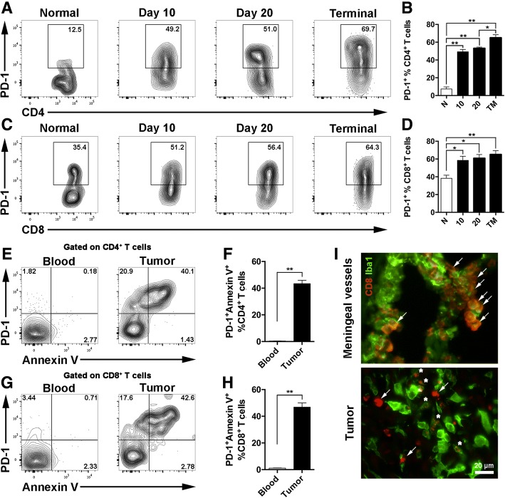
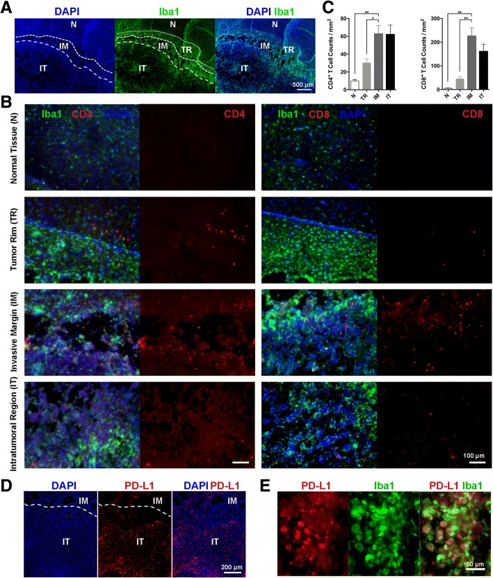
The distribution of tumor-infiltrating T cells and PD-L1 expression was analyzed via immunofluorescence in orthotopic murine glioma model. The expression of PD-L1 in immune cell populations was detected by flow cytometry. Data excavated from TCGA LGG/GBM datasets and the Ivy Glioblastoma Atlas Project was used for in silico analysis of the correlation among genes and survival.
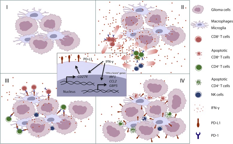
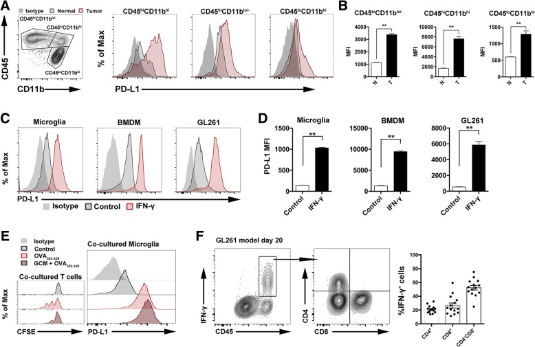
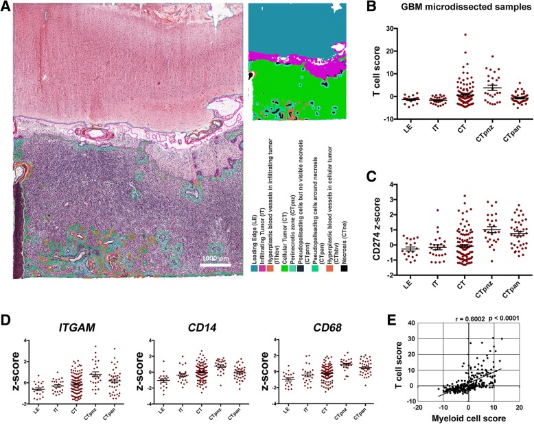
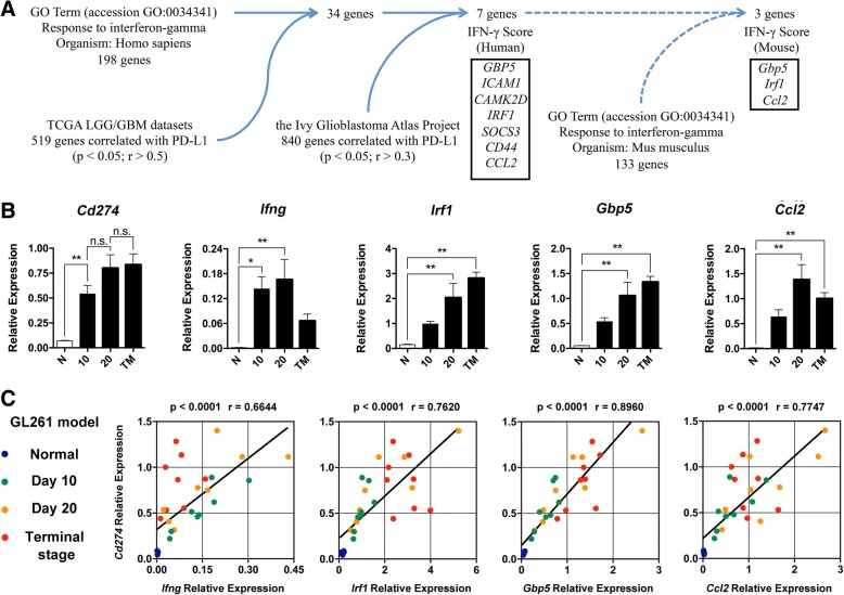
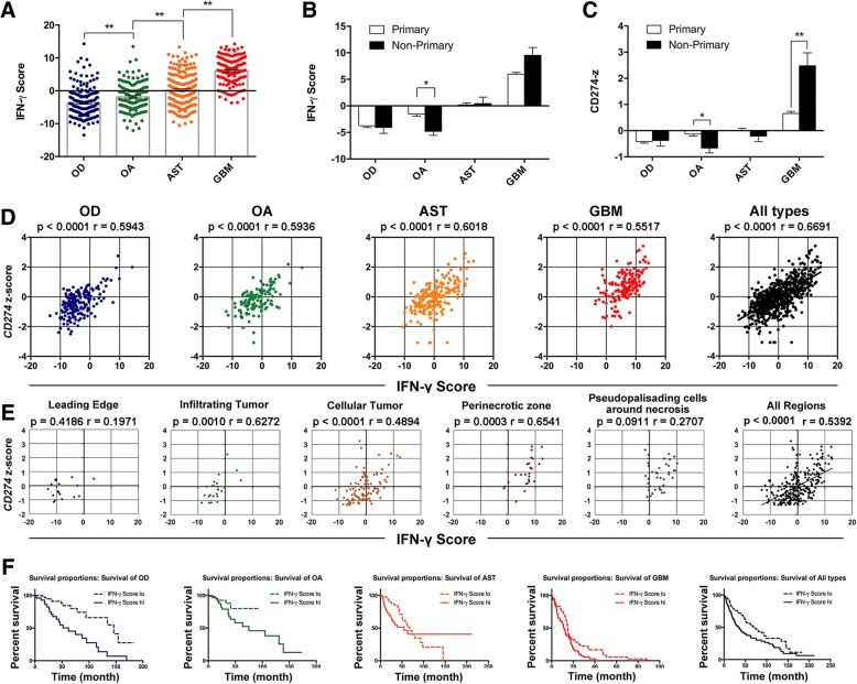
The distribution of tumor-infiltrating T cells and PD-L1 expression, which parallels in murine orthotopic glioma model and human glioma microdissections, was interrelated. The IFN-γ level was positively correlated with PD-L1 expression in murine glioma. Further, IFN-γ induces PD-L1 expression on primary cultured microglia, bone marrow-derived macrophages, and GL261 glioma cells in vitro. Seven IFN-γ-induced genes, namely GBP5, ICAM1, CAMK2D, IRF1, SOCS3, CD44, and CCL2, were selected to calculate as substitute indicator for IFN-γ level. By combining the relative expression of the listed IFN-γ-induced genes, IFN-γ score was positively correlated with PD-L1 expression in different anatomic structures of human glioma and in glioma of different malignancies.
Our study identified the distribution of tumor-infiltrating T cells and PD-L1 expression in murine glioma model and human glioma samples. And we found that IFN-γ is an important cause of PD-L1 expression in the glioma microenvironment. Further, we proposed IFN-γ score aggregated from the expressions of the listed IFN-γ-induced genes as a complementary prognostic indicator for anti-PD-1/PD-L1 therapy.
PD-L1 是一种免疫抑制性受体配体,通过与受体 PD-1 结合导致 T 细胞功能障碍和凋亡,而 PD-1 则在制动炎症反应和共谋肿瘤免疫逃逸方面发挥作用。然而,在神经胶质瘤中,肿瘤微环境中 PD-L1 表达的原因尚不清楚。此外,迫切需要辅助生物标志物来筛选可能对抗 PD-1/PD-L1 治疗有反应的神经胶质瘤患者。
通过免疫荧光分析原位鼠神经胶质瘤模型中肿瘤浸润性 T 细胞和 PD-L1 表达的分布。通过流式细胞术检测免疫细胞群中 PD-L1 的表达。利用 TCGA LGG/GBM 数据集和 Ivy 神经胶质瘤图谱项目的数据进行基因与生存之间相关性的计算机分析。
在鼠原位神经胶质瘤模型和人神经胶质瘤微切片中,肿瘤浸润性 T 细胞的分布和 PD-L1 表达相互关联。IFN-γ 水平与鼠神经胶质瘤中 PD-L1 的表达呈正相关。此外,IFN-γ 在体外诱导原代培养的小胶质细胞、骨髓来源的巨噬细胞和 GL261 神经胶质瘤细胞表达 PD-L1。选择 7 个 IFN-γ 诱导基因(GBP5、ICAM1、CAMK2D、IRF1、SOCS3、CD44 和 CCL2)计算作为 IFN-γ 水平的替代指标。通过组合列出的 IFN-γ 诱导基因的相对表达,IFN-γ 评分与不同解剖结构的人神经胶质瘤和不同恶性程度的神经胶质瘤中的 PD-L1 表达呈正相关。
本研究鉴定了鼠神经胶质瘤模型和人神经胶质瘤样本中肿瘤浸润性 T 细胞的分布和 PD-L1 表达。我们发现 IFN-γ 是肿瘤微环境中 PD-L1 表达的重要原因。进一步提出,从列出的 IFN-γ 诱导基因的表达中聚合的 IFN-γ 评分作为抗 PD-1/PD-L1 治疗的补充预后指标。