Schachtele Scott J, Hu Shuxian, Sheng Wen S, Mutnal Manohar B, Lokensgard James R
Department of Medicine, Center for Infectious Diseases and Microbiology Translational Research, University of Minnesota, McGuire Translational Research Facility, Minneapolis, Minnesota.
Glia. 2014 Oct;62(10):1582-94. doi: 10.1002/glia.22701. Epub 2014 Jun 3.
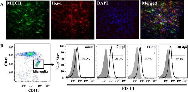
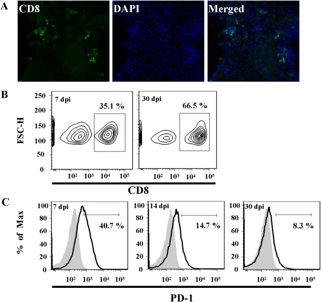
Engagement of the programmed death (PD)-1 receptor on activated cells by its ligand (PD-L1) is a mechanism for suppression of activated T-lymphocytes. Microglia, the resident inflammatory cells of the brain, are important for pathogen detection and initiation of innate immunity, however, a novel role for these cells as immune regulators has also emerged. PD-L1 on microglia has been shown to negatively regulate T-cell activation in models of multiple sclerosis and acute viral encephalitis. In this study, we investigated the role of glial cell PD-L1 in controlling encephalitogenic CD8(+) T-lymphocytes, which infiltrate the brain to manage viral infection, but remain to produce chronic neuroinflammation. Using a model of chronic neuroinflammation following murine cytomegalovirus (MCMV)-induced encephalitis, we found that CD8(+) T-cells persisting within the brain expressed PD-1. Conversely, activated microglia expressed PD-L1. In vitro, primary murine microglia, which express low basal levels of PD-L1, upregulated the co-inhibitory ligand on IFN-γ-treatment. Blockade of the PD-1: PD-L1 pathway in microglial: CD8(+) T-cell co-cultures increased T-cell IFN-γ and interleukin (IL)-2 production. We observed a similar phenomenon following blockade of this co-inhibitory pathway in astrocyte: CD8(+) T-cell co-cultures. Using ex vivo cultures of brain leukocytes, including microglia and CD8(+) T-cells, obtained from mice with MCMV-induced chronic neuroinflammation, we found that neutralization of either PD-1 or PD-L1 increased IFN-γ production from virus-specific CD8(+) T-cells stimulated with MCMV IE1168-176 peptide. These data demonstrate that microglia and astrocytes control antiviral T-cell responses and suggest a therapeutic potential of PD1: PD-L1 modulation to manage the deleterious consequences of uncontrolled neuroinflammation.
其配体(PD-L1)与活化细胞上的程序性死亡(PD)-1受体结合是抑制活化T淋巴细胞的一种机制。小胶质细胞是大脑中的常驻炎症细胞,对病原体检测和先天免疫的启动很重要,然而,这些细胞作为免疫调节因子的新作用也已显现。在多发性硬化症和急性病毒性脑炎模型中,小胶质细胞上的PD-L1已被证明对T细胞活化具有负调节作用。在本研究中,我们调查了神经胶质细胞PD-L1在控制致脑炎性CD8(+) T淋巴细胞中的作用,这些细胞浸润大脑以应对病毒感染,但仍会引发慢性神经炎症。使用鼠巨细胞病毒(MCMV)诱导的脑炎后慢性神经炎症模型,我们发现持续存在于大脑中的CD8(+) T细胞表达PD-1。相反,活化的小胶质细胞表达PD-L1。在体外,表达低基础水平PD-L1的原代鼠小胶质细胞在IFN-γ处理后上调了共抑制配体。在小胶质细胞与CD8(+) T细胞共培养中阻断PD-1:PD-L1途径可增加T细胞IFN-γ和白细胞介素(IL)-2的产生。在星形胶质细胞与CD8(+) T细胞共培养中阻断这种共抑制途径后,我们观察到了类似现象。使用从患有MCMV诱导的慢性神经炎症的小鼠获得的包括小胶质细胞和CD8(+) T细胞在内的脑白细胞的离体培养物,我们发现中和PD-1或PD-L1可增加用MCMV IE1168-176肽刺激的病毒特异性CD8(+) T细胞的IFN-γ产生。这些数据表明小胶质细胞和星形胶质细胞可控制抗病毒T细胞反应,并提示PD1:PD-L1调节在管理不受控制的神经炎症的有害后果方面具有治疗潜力。