Translational and Molecular Imaging Institute, Icahn School of Medicine at Mount Sinai, New York, New York; Department of Medical Biochemistry, Academic Medical Center, Amsterdam, the Netherlands.
In Vivo Cellular and Molecular Imaging Laboratory, Vrije Universiteit Brussel, Brussels, Belgium.
JACC Cardiovasc Imaging. 2019 Oct;12(10):2015-2026. doi: 10.1016/j.jcmg.2018.07.027. Epub 2018 Oct 17.
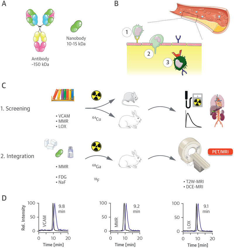
This study sought to develop an integrative positron emission tomography (PET) with magnetic resonance imaging (MRI) procedure for accurate atherosclerotic plaque phenotyping, facilitated by clinically approved and nanobody radiotracers.
Noninvasive characterization of atherosclerosis remains a challenge in clinical practice. The limitations of current diagnostic methods demonstrate that, in addition to atherosclerotic plaque morphology and composition, disease activity needs to be evaluated.
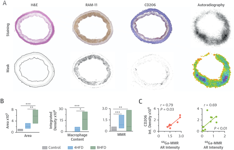
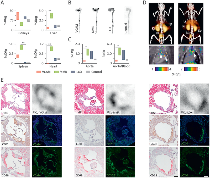
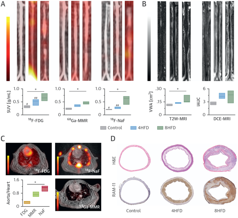
We screened 3 nanobody radiotracers targeted to different biomarkers of atherosclerosis progression, namely vascular cell adhesion molecule (VCAM)-1, lectin-like oxidized low-density lipoprotein receptor (LOX)-1, and macrophage mannose receptor (MMR). The nanobodies, initially radiolabeled with copper-64 (Cu), were extensively evaluated in Apoe mice and atherosclerotic rabbits using a combination of in vivo PET/MRI readouts and ex vivo radioactivity counting, autoradiography, and histological analyses.
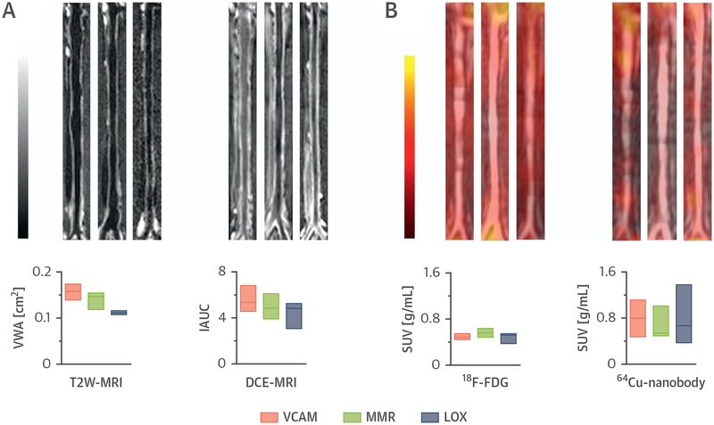
The 3 nanobody radiotracers accumulated in atherosclerotic plaques and displayed short circulation times due to fast renal clearance. The MMR nanobody was selected for labeling with gallium-68 (Ga), a short-lived radioisotope with high clinical relevance, and used in an ensuing atherosclerosis progression PET/MRI study. Macrophage burden was longitudinally studied by Ga-MMR-PET, plaque burden by T2-weighted MRI, and neovascularization by dynamic contrast-enhanced (DCE) MRI. Additionally, inflammation and microcalcifications were evaluated by fluorine-18 (F)-labeled fluorodeoxyglucose (F-FDG) and F-sodium fluoride (F-NaF) PET, respectively. We observed an increase in all the aforementioned measures as disease progressed, and the imaging signatures correlated with histopathological features.
We have evaluated nanobody-based radiotracers in rabbits and developed an integrative PET/MRI protocol that allows noninvasive assessment of different processes relevant to atherosclerosis progression. This approach allows the multiparametric study of atherosclerosis and can aid in early stage anti-atherosclerosis drug trials.
本研究旨在开发一种整合正电子发射断层扫描(PET)与磁共振成像(MRI)的方法,以利用临床批准的纳米体放射性示踪剂准确地对动脉粥样硬化斑块进行表型分析。
在临床实践中,非侵入性地对动脉粥样硬化进行特征描述仍然是一个挑战。目前诊断方法的局限性表明,除了动脉粥样硬化斑块的形态和组成外,还需要评估疾病的活动程度。
我们筛选了 3 种针对动脉粥样硬化进展不同生物标志物的纳米体放射性示踪剂,即血管细胞黏附分子(VCAM)-1、凝集素样氧化低密度脂蛋白受体(LOX)-1 和巨噬细胞甘露糖受体(MMR)。最初用铜-64(Cu)对这些纳米体进行放射性标记,然后在载脂蛋白 E (Apoe)小鼠和动脉粥样硬化兔中进行了广泛的评估,采用了结合体内 PET/MRI 读出和体外放射性计数、放射自显影和组织学分析的方法。
这 3 种纳米体放射性示踪剂在动脉粥样硬化斑块中积累,并由于快速的肾脏清除而表现出短的循环时间。由于镓-68(Ga)是一种半衰期短且具有高临床相关性的放射性同位素,因此选择 MMR 纳米体进行标记,并将其用于随后的动脉粥样硬化进展 PET/MRI 研究。通过 Ga-MMR-PET 研究巨噬细胞负荷,通过 T2 加权 MRI 研究斑块负荷,通过动态对比增强(DCE)MRI 研究新生血管化。此外,通过氟-18(F)标记的氟脱氧葡萄糖(F-FDG)和 F-氟化钠(F-NaF)PET 分别评估炎症和微钙化。随着疾病的进展,我们观察到所有上述指标都增加了,并且成像特征与组织病理学特征相关。
我们在兔中评估了基于纳米体的放射性示踪剂,并开发了一种整合的 PET/MRI 方案,该方案允许对与动脉粥样硬化进展相关的不同过程进行非侵入性评估。这种方法允许对动脉粥样硬化进行多参数研究,并有助于早期抗动脉粥样硬化药物试验。