OMFS-IMPATH Research Group, Department of Imaging and Pathology, Katholieke Universiteit Leuven.
DentoMaxillofacial Radiology Center, University Hospitals Leuven, Leuven, Belgium.
Eur J Orthod. 2019 Aug 8;41(4):381-389. doi: 10.1093/ejo/cjy066.
Taking into account radiation doses, safety, and protection, we highlighted the features in which cone-beam computed tomography (CBCT) can offer an advantage compared to the conventional two-dimensional imaging in paediatric dentistry before orthodontic treatment.
The aim of this article was to conduct a systematic review to assess the diagnostic efficacy of CBCT in the paediatric population at a pre-orthodontic phase.
MEDLINE via PubMed was searched to identify all peer-reviewed articles potentially relevant to the review until 1 July 2018. Relevant publications were selected by two reviewers independently.
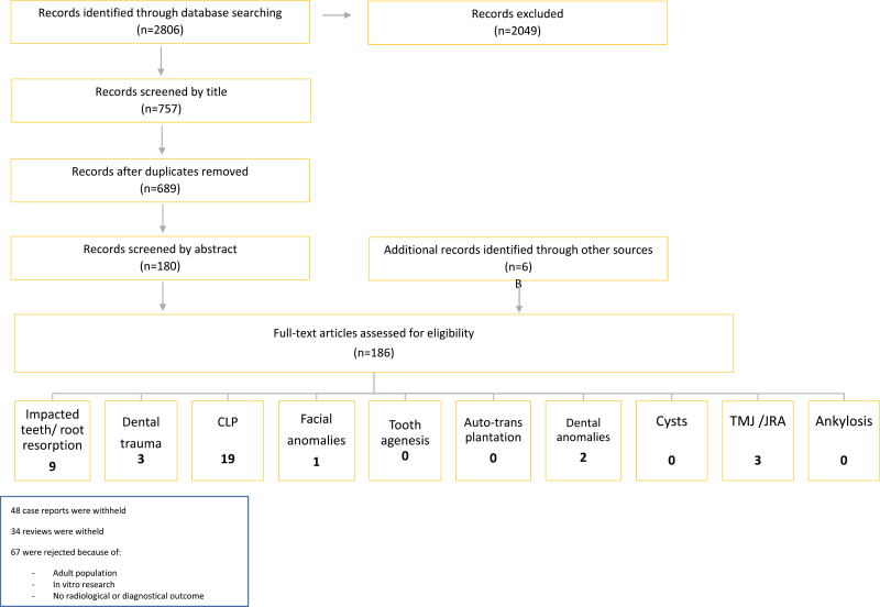
The literature selection for this systematic review was carried out according to the Preferred Reporting Items for Systematic Reviews and Meta-Analyses (PRISMA) statement and was based on predetermined inclusion criteria.
Data were collected on overall study characteristics and examination characteristics of the selected studies. Methodological quality of the selected studies was evaluated. Original studies were assessed using the Quality Assessment of Diagnostic Accuracy Studies (QUADAS) tool. Thereafter, levels of evidence were obtained according to Grading of Recommendations Assessment, Development and Evaluation criteria.
As a result of the QUADAS assessment, a total of 37 articles were included in the protocol. Following a proper protocol, CBCT was regarded as a reliable tool for assessment and management of impacted canine and root fracture. It provided a better evaluation of normal and pathological condylar shape and volume. CBCT was a superior choice for pre-surgical diagnostic applications in cleft lip and/or palate over a medical computed tomography based on its lower radiation exposure, shorter investigation time, and low purchase costs.
CBCT is justified only in those cases where conventional radiography fails to provide a correct diagnosis of pathology. Therefore, it cannot be regarded as a standard method of diagnosis. CBCT imaging may also be justified when it positively affects treatment options or provides treatment optimization.
None.
None to declare.
考虑到辐射剂量、安全性和防护,我们强调了锥形束计算机断层扫描(CBCT)相对于传统二维成像在儿童牙科正畸治疗前的优势。
本文旨在进行系统评价,以评估 CBCT 在儿童正畸前阶段的诊断效能。
通过 MEDLINE 下的 PubMed 检索,检索截至 2018 年 7 月 1 日所有可能与综述相关的同行评审文献。由两名评审员独立选择相关出版物。
本系统评价的文献选择按照系统评价和荟萃分析的首选报告项目(PRISMA)声明进行,并基于预定的纳入标准。
收集了所选研究的总体研究特征和检查特征的数据。评估了所选研究的方法学质量。使用诊断准确性研究的质量评估(QUADAS)工具评估原始研究。然后,根据推荐评估、制定和评价标准获得证据水平。
根据 QUADAS 评估,共有 37 篇文章纳入方案。按照适当的方案,CBCT 被认为是评估和管理埋伏牙和根折的可靠工具。它提供了对正常和病理髁突形状和体积的更好评估。与基于医学 CT 的术前诊断应用相比,CBCT 由于辐射暴露量较低、检查时间较短和购买成本较低,因此是唇裂和/或腭裂术前诊断的首选。
只有在常规放射学不能正确诊断疾病的情况下,才可以使用 CBCT。因此,它不能被视为一种标准的诊断方法。当 CBCT 对治疗方案有积极影响或提供治疗优化时,也可以使用 CBCT 成像。
无。
无。