From the Department of Neurosurgery (M.I., M.A., S.I., Z.C., E.H.W., S.L.L., D.L.S., M.Y.C., G.K.S.).
Department of Pediatrics (A.G.L.).
Stroke. 2018 Sep;49(9):2191-2199. doi: 10.1161/STROKEAHA.118.021508.
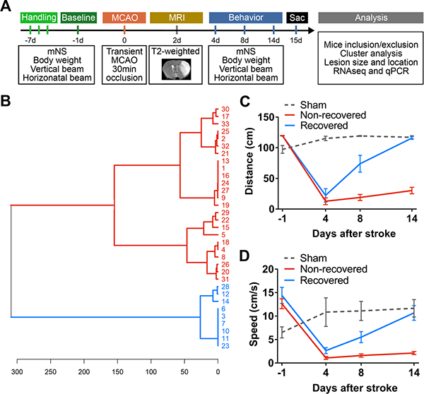
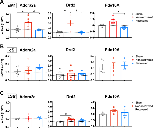
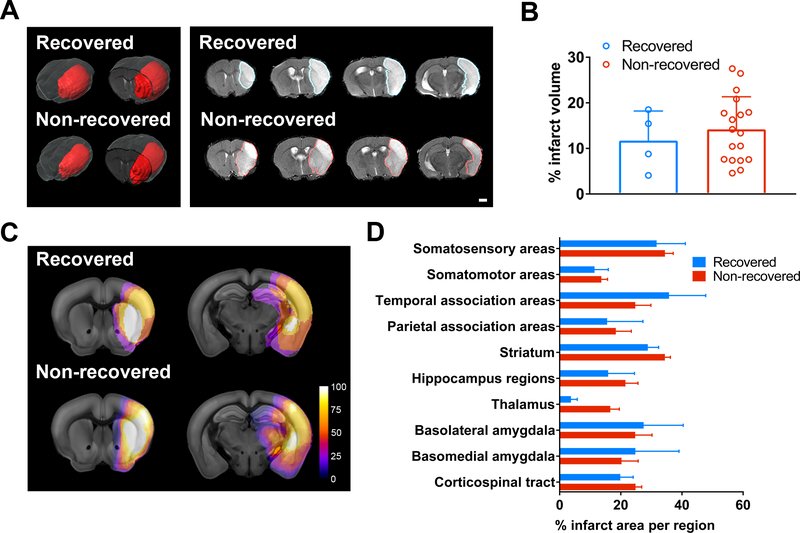
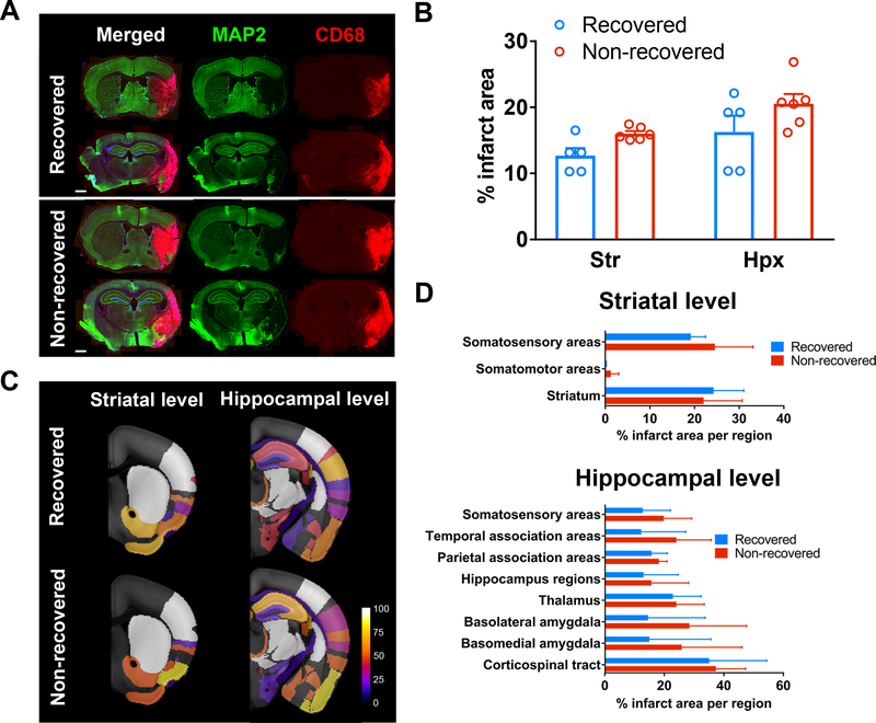
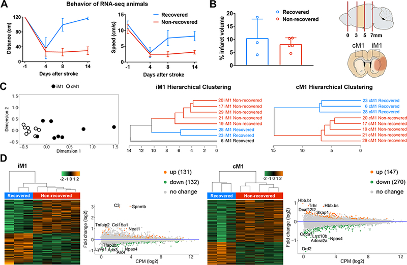
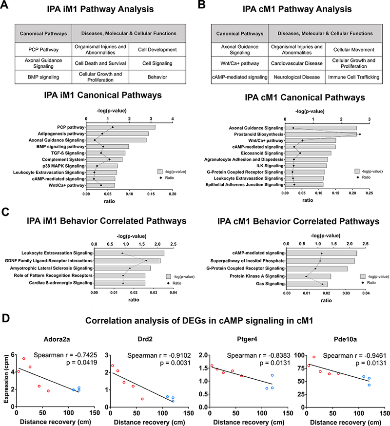
Background and Purpose- Many restorative therapies have been used to study brain repair after stroke. These therapeutic-induced changes have revealed important insights on brain repair and recovery mechanisms; however, the intrinsic changes that occur in spontaneously recovery after stroke is less clear. The goal of this study is to elucidate the intrinsic changes in spontaneous recovery after stroke, by directly investigating the transcriptome of primary motor cortex in mice that naturally recovered after stroke. Methods- Male C57BL/6J mice were subjected to transient middle cerebral artery occlusion. Functional recovery was evaluated using the horizontal rotating beam test. A novel in-depth lesion mapping analysis was used to evaluate infarct size and locations. Ipsilesional and contralesional primary motor cortices (iM1 and cM1) were processed for RNA-sequencing transcriptome analysis. Results- Cluster analysis of the stroke mice behavior performance revealed 2 distinct recovery groups: a spontaneously recovered and a nonrecovered group. Both groups showed similar lesion profile, despite their differential recovery outcome. RNA-sequencing transcriptome analysis revealed distinct biological pathways in the spontaneously recovered stroke mice, in both iM1 and cM1. Correlation analysis revealed that 38 genes in the iM1 were significantly correlated with improved recovery, whereas 74 genes were correlated in the cM1. In particular, ingenuity pathway analysis highlighted the involvement of cAMP signaling in the cM1, with selective reduction of Adora2a (adenosine receptor A2A), Drd2 (dopamine receptor D2), and Pde10a (phosphodiesterase 10A) expression in recovered mice. Interestingly, the expressions of these genes in cM1 were negatively correlated with behavioral recovery. Conclusions- Our RNA-sequencing data revealed a panel of recovery-related genes in the motor cortex of spontaneously recovered stroke mice and highlighted the involvement of contralesional cortex in spontaneous recovery, particularly Adora2a, Drd2, and Pde10a-mediated cAMP signaling pathway. Developing drugs targeting these candidates after stroke may provide beneficial recovery outcome.
背景与目的- 许多修复疗法已被用于研究中风后的大脑修复。这些治疗引起的变化揭示了重要的关于大脑修复和恢复机制的见解;然而,中风后自然恢复过程中发生的内在变化尚不清楚。本研究的目的是通过直接研究中风后自然恢复的小鼠初级运动皮层的转录组,阐明中风后自发恢复的内在变化。方法- 雄性 C57BL/6J 小鼠接受短暂性大脑中动脉闭塞。使用水平旋转梁测试评估功能恢复。使用新的深入损伤映射分析评估梗死大小和位置。对同侧和对侧初级运动皮层(iM1 和 cM1)进行 RNA 测序转录组分析。结果- 对中风小鼠行为表现的聚类分析显示出 2 个不同的恢复组:自发恢复组和未恢复组。尽管恢复结果不同,但两组的损伤情况相似。RNA 测序转录组分析显示,在自发恢复的中风小鼠中,iM1 和 cM1 中存在不同的生物学途径。相关性分析显示,iM1 中的 38 个基因与改善恢复显著相关,而 cM1 中有 74 个基因相关。特别是,通路分析突出了 cAMP 信号通路在 cM1 中的作用,在恢复的小鼠中,Adora2a(腺苷受体 A2A)、Drd2(多巴胺受体 D2)和 Pde10a(磷酸二酯酶 10A)的表达选择性降低。有趣的是,cM1 中这些基因的表达与行为恢复呈负相关。结论- 我们的 RNA 测序数据揭示了一组与自发性中风恢复的运动皮层相关的基因,并强调了对侧皮层在自发性恢复中的作用,特别是 Adora2a、Drd2 和 Pde10a 介导的 cAMP 信号通路。中风后针对这些候选药物的开发可能会提供有益的恢复效果。