Department of Human Genetics, University of Pittsburgh, Pittsburgh, PA, USA.
Department of Pediatrics, Children's Hospital of Pittsburgh of UPMC, University of Pittsburgh, Pittsburgh, PA, USA.
Mol Psychiatry. 2021 Jan;26(1):309-321. doi: 10.1038/s41380-018-0246-7. Epub 2018 Oct 25.
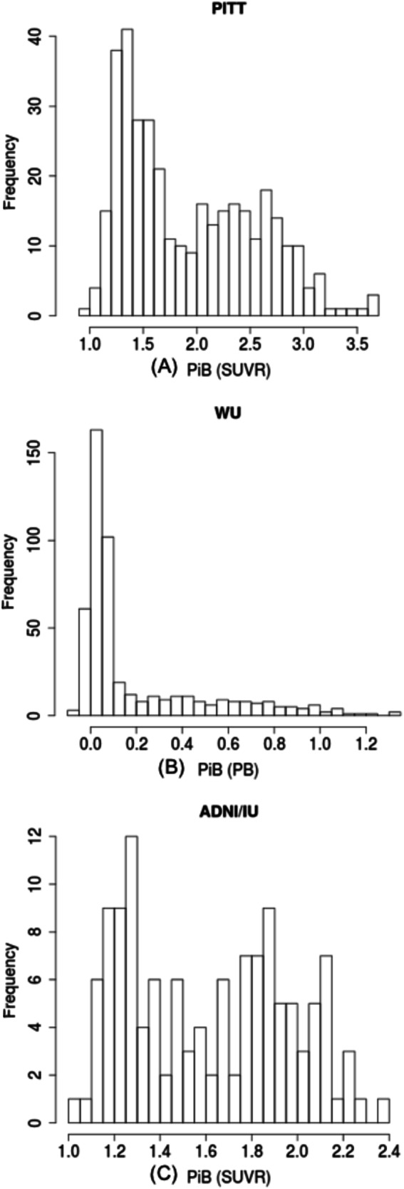
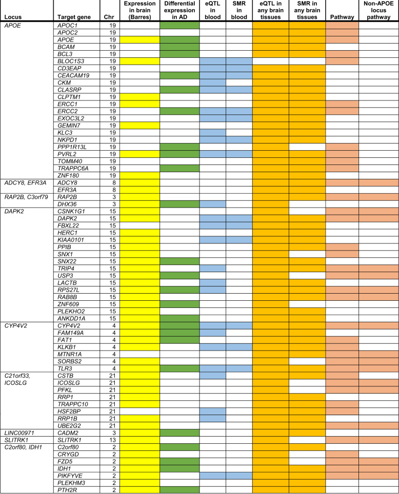
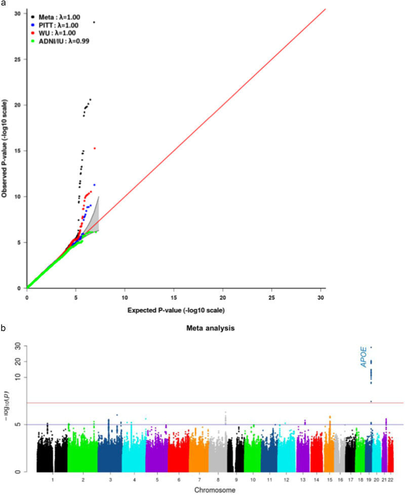
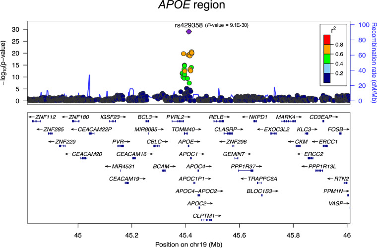
Deposition of amyloid plaques in the brain is one of the two main pathological hallmarks of Alzheimer's disease (AD). Amyloid positron emission tomography (PET) is a neuroimaging tool that selectively detects in vivo amyloid deposition in the brain and is a reliable endophenotype for AD that complements cerebrospinal fluid biomarkers with regional information. We measured in vivo amyloid deposition in the brains of ~1000 subjects from three collaborative AD centers and ADNI using C-labeled Pittsburgh Compound-B (PiB)-PET imaging followed by meta-analysis of genome-wide association studies, first to our knowledge for PiB-PET, to identify novel genetic loci for this endophenotype. The APOE region showed the most significant association where several SNPs surpassed the genome-wide significant threshold, with APOE4 being most significant (P-meta = 9.09E-30; β = 0.18). Interestingly, after conditioning on APOE4, 14 SNPs remained significant at P < 0.05 in the APOE region that were not in linkage disequilibrium with APOE4. Outside the APOE region, the meta-analysis revealed 15 non-APOE loci with P < 1E-05 on nine chromosomes, with two most significant SNPs on chromosomes 8 (P-meta = 4.87E-07) and 3 (P-meta = 9.69E-07). Functional analyses of these SNPs indicate their potential relevance with AD pathogenesis. Top 15 non-APOE SNPs along with APOE4 explained 25-35% of the amyloid variance in different datasets, of which 14-17% was explained by APOE*4 alone. In conclusion, we have identified novel signals in APOE and non-APOE regions that affect amyloid deposition in the brain. Our data also highlights the presence of yet to be discovered variants that may be responsible for the unexplained genetic variance of amyloid deposition.
淀粉样斑块在大脑中的沉积是阿尔茨海默病(AD)的两个主要病理标志之一。淀粉样蛋白正电子发射断层扫描(PET)是一种神经影像学工具,可选择性地检测大脑中的体内淀粉样蛋白沉积,是 AD 的可靠内表型,与脑脊液生物标志物相结合,提供了区域信息。我们使用 C 标记的匹兹堡化合物-B(PiB)-PET 成像测量了来自三个合作 AD 中心和 ADNI 的约 1000 名受试者的大脑中的体内淀粉样蛋白沉积,然后对全基因组关联研究进行荟萃分析,这是首次针对 PiB-PET 进行的分析,以确定该内表型的新遗传位点。APOE 区域显示出最显著的关联,其中几个 SNP 超过了全基因组显著阈值,APOE4 最显著(P-荟萃分析 = 9.09E-30;β = 0.18)。有趣的是,在 APOE4 条件下,APOE 区域内还有 14 个 SNP 仍然显著(P<0.05),与 APOE4 没有连锁不平衡。在 APOE 区域之外,荟萃分析显示了 15 个非 APOE 基因座,在 9 条染色体上有 15 个 SNP 达到 P<1E-05,其中两个最显著的 SNP 位于 8 号染色体(P-荟萃分析 = 4.87E-07)和 3 号染色体(P-荟萃分析 = 9.69E-07)。这些 SNP 的功能分析表明了它们与 AD 发病机制的潜在相关性。前 15 个非 APOE SNP 加上 APOE4 可以解释不同数据集 25-35%的淀粉样蛋白变异,其中 14-17%由 APOE*4 单独解释。总之,我们已经在 APOE 和非 APOE 区域中确定了新的信号,这些信号会影响大脑中的淀粉样蛋白沉积。我们的数据还突出了存在尚未发现的变体,这些变体可能负责淀粉样蛋白沉积的遗传变异未被解释的原因。