Danish Colorectal Cancer Center South, Vejle Hospital, Part of Lillebaelt Hospital, Beriderbakken 4, 7100, Vejle, Denmark.
Department of Clinical Pathology, Vejle Hospital, Part of Lillebaelt Hospital, Beriderbakken 4, 7100, Vejle, Denmark.
Clin Exp Metastasis. 2018 Dec;35(8):819-830. doi: 10.1007/s10585-018-9945-3. Epub 2018 Oct 25.
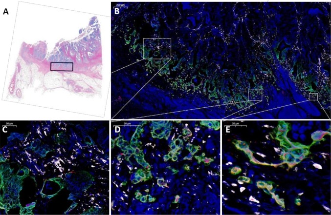
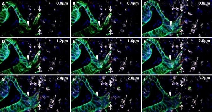
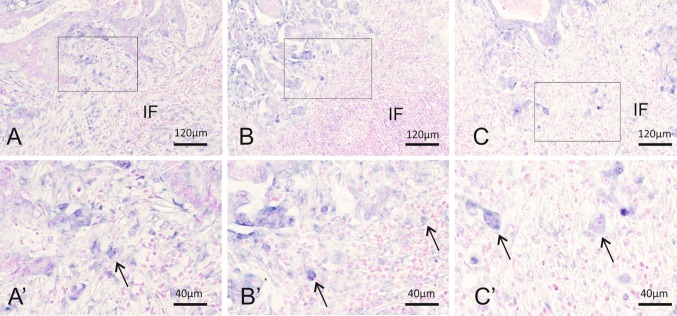
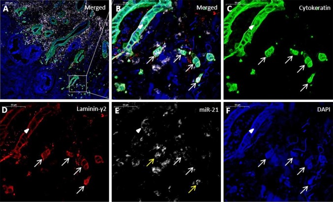
MicroRNA-21 (miR-21) expression in stromal fibroblastic cells in colorectal cancer is well-documented, whereas miR-21 expression in tumor budding cells (TBCs) is poorly described. TBCs are locally invasive carcinoma cells with increased metastatic properties and characteristics of epithelial to mesenchymal transition. This study was conducted to better characterize the expression of miR-21 in TBCs. First, chromogenic miR-21 in situ hybridization (ISH) staining was performed in 58 colon adenocarcinomas with evident TBCs. Then, to obtain unambiguous identification of miR-21 in the TBCs, twenty cases were selected for an additional multiplex fluorescence analysis combining miR-21 ISH with cytokeratin and laminin-5γ2 immunofluorescence. Employing confocal slide scanning microscopy, comprehensive digital images of the invasive front (10-40 mm) were obtained from 16 of the 20 cases, and miR-21 expression was evaluated in cytokeratin-positive TBCs. The high resolution of the confocal digital slide images allowed a detailed examination of the confocal stacks of the multiplex-stained tissue sections. The cases with the highest fraction of miR-21 positive TBCs were all stage III cancers defined by the presence of regional lymph node metastasis. Some of the miR-21 positive TBCs were also laminin-5γ2 positive. The confocal image stacks also revealed that some TBCs were actually directly connected to malignant glands. In conclusion, miR-21 expression was unambiguously identified in TBCs by evaluation of digital slides obtained by confocal slide scanning microscopy. In addition, the digital confocal slides provided a more detailed understanding of local cancer cell invasion by allowing evaluation of the cell structures in three dimensions.
微 RNA-21(miR-21)在结直肠癌细胞中的表达已有充分的文献记载,而肿瘤芽细胞(TBC)中的 miR-21 表达则描述甚少。TBC 是具有增加的转移性特性和上皮到间充质转化特征的局部侵袭性癌细胞。本研究旨在更好地描述 TBC 中 miR-21 的表达。首先,在 58 例明显有 TBC 的结肠腺癌中进行了显色 miR-21 原位杂交(ISH)染色。然后,为了明确鉴定 TBC 中的 miR-21,选择了 20 例进行结合 miR-21 ISH 与细胞角蛋白和层粘连蛋白-5γ2 免疫荧光的多重荧光分析。采用共聚焦幻灯片扫描显微镜,从 20 例中的 16 例获得了侵袭前沿(10-40mm)的综合数字图像,并在细胞角蛋白阳性的 TBC 中评估了 miR-21 的表达。共聚焦数字幻灯片图像的高分辨率允许对多染组织切片的共聚焦堆栈进行详细检查。miR-21 阳性 TBC 比例最高的病例均为存在区域淋巴结转移的 III 期癌症。一些 miR-21 阳性 TBC 也为层粘连蛋白-5γ2 阳性。共聚焦图像堆栈还揭示了一些 TBC 实际上直接与恶性腺体相连。总之,通过共聚焦幻灯片扫描显微镜获得的数字幻灯片评估,明确鉴定了 TBC 中的 miR-21 表达。此外,数字共聚焦幻灯片通过评估三维细胞结构,提供了对局部癌细胞侵袭的更详细理解。