Zainuri Masagus, Putri Ratih Rinendya, Bachtiar Endang W
Center for Research and Development of Biomedical and Basic Health Technology, National Institute Health Research and Development, Ministry of Health Republic of Indonesia, Jakarta, Indonesia.
Faculty of Dentistry, Department of Oral Biology, Oral Sciences Research Center, Universitas Indonesia, Jakarta, Indonesia.
Interv Med Appl Sci. 2018 Mar;10(1):33-37. doi: 10.1556/1646.10.2018.06.
This study aims to establish the isolation method of stem cells from pulp tissue of carious deciduous teeth.
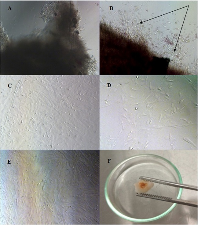
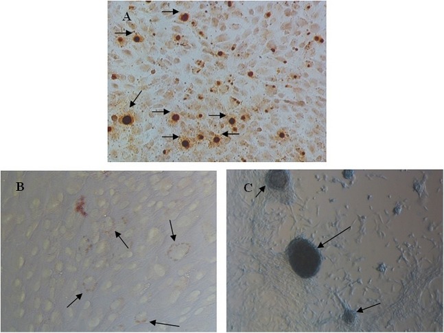
The teeth were soaked in 1% povidone-iodine solution for about 1 min followed by washing in PBS with 1% antibiotic-antimycotic thrice. Dental pulp tissue was removed by extirpation, and then cultivated in the culture medium. Characterization of mesenchymal stem cell (MSC) was carried out using human MSC analysis kit with positive markers CD90, CD73, and CD105, but negative for expressions of CD45, CD34, CD11b, CD19, and HLA-DR. Differentiation capacity of stem cells from human exfoliated deciduous (SHED) was determined by staining with Alizarin S, Alcian Blue, and Oil Red O.
There is no contamination after 3 days of culture. SHED derived from dental pulp were expressions of 99.2% of positive marker and 0.3% of the negative marker. At passage 5, SHED was differentiated into osteocyte, chondrocyte, and adipocyte types of cells in the induction medium.
SHED derived from carious deciduous teeth can be used as a source of stem cell for regenerative medicine.
本研究旨在建立从龋损乳牙牙髓组织中分离干细胞的方法。
将牙齿浸泡于1%聚维酮碘溶液中约1分钟,然后用含1%抗生素 - 抗真菌剂的磷酸盐缓冲盐水(PBS)冲洗三次。通过摘除术去除牙髓组织,然后在培养基中培养。使用人间充质干细胞分析试剂盒对间充质干细胞(MSC)进行鉴定,其阳性标志物为CD90、CD73和CD105,而CD45、CD34、CD11b、CD19和HLA - DR表达为阴性。通过茜素红S、阿尔辛蓝和油红O染色来确定人脱落乳牙干细胞(SHED)的分化能力。
培养3天后无污染。牙髓来源的SHED阳性标志物表达率为99.2%,阴性标志物表达率为0.3%。在第5代时,SHED在诱导培养基中分化为骨细胞、软骨细胞和脂肪细胞类型。
龋损乳牙来源的SHED可作为再生医学的干细胞来源。