Garcia-Pradas Lidia, Gleiser Corinna, Wizenmann Andrea, Wolburg Hartwig, Mack Andreas F
Institut für klinische Anatomie und Zellanalytik, Universität Tübingen, Tübingen, Germany.
Institut für Pathologie und Neuropathologie, Universität Tübingen, Tübingen, Germany.
Front Mol Neurosci. 2018 Oct 10;11:367. doi: 10.3389/fnmol.2018.00367. eCollection 2018.
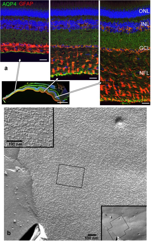
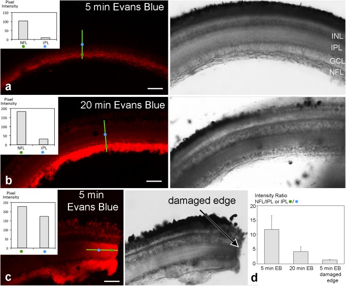
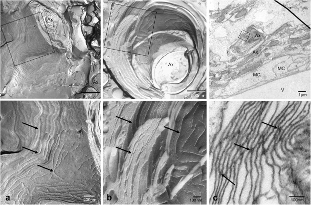
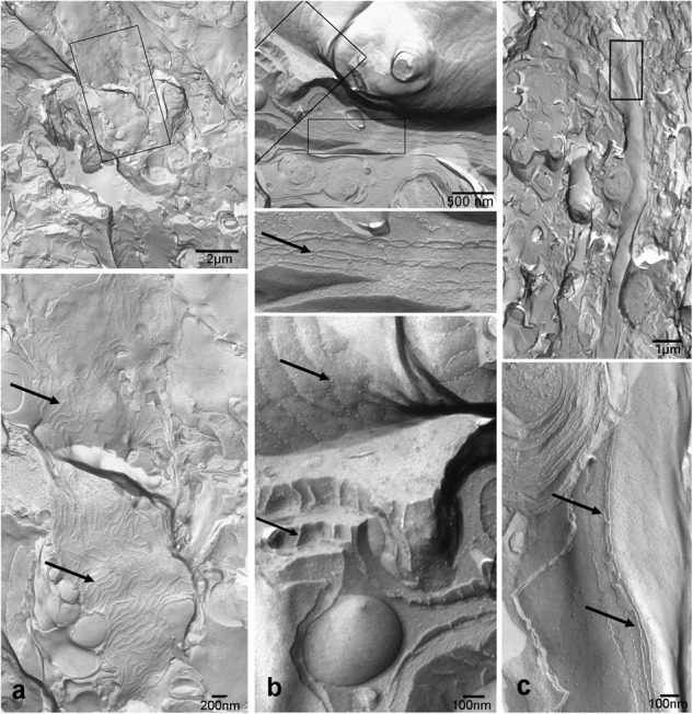
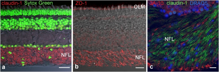
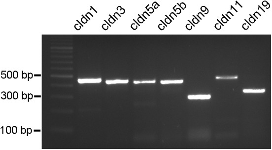
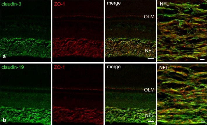
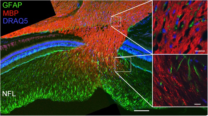
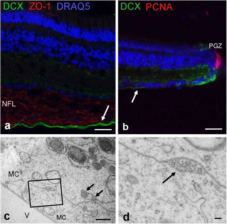
In the retina of teleost fish, cell addition continues throughout life involving proliferation and axonal growth. To study how this is achieved in a fully functioning retina, we investigated the nerve fiber layer (NFL) of the cichlid fish for components that might regulate the extracellular environment. We hypothesized that growing axons are surrounded by different cell structures than signal conducting axons. Using immunohistochemistry and freeze fracture electron microscopy we found that the endfeet of Müller cells (MCs) expressed aquaporin-4 but not in high densities as in mammals. The presence of this water channel indicates the involvement of MCs in water homeostasis. Remarkably, we discovered conspicuous tight junctions in the retinal NFL. These tight junctions formed branching strands between myelin-like wrappings of ganglion cell axons that differed morphologically from any known myelin, and also an elaborate meshwork on large membrane faces between axons. We speculated that these tight junctions have additional functions than solely facilitating nerve conductance. Immunostainings against the adaptor protein ZO-1 labeled the NFL as did antibodies against the mammalian claudin-1, 3, and 19. Performing PCR analysis, we showed expression of claudin-1, 3, 5a, 5b, 9, 11, and 19 in the fish retina, claudins that typically occur at brain barriers or myelin. We could show by immunostains for doublecortin, a marker for differentiating neurons, that new axons are not surrounded by the myelin-like wrappings but only by the endfeet of MCs. We hypothesize that the tight junctions in the NFL of fish might contribute to the separation of an extracellular space around axons facilitating conductance, from a growth-promoting environment. For a functional test we applied Evans Blue dye to eye cup preparations which showed a retention of the dye in the NFL. This indicates that these remarkable tight junctions can indeed act as a diffusion barrier.
在硬骨鱼的视网膜中,细胞增殖和轴突生长持续一生。为了研究在功能完备的视网膜中这一过程是如何实现的,我们研究了丽鱼科鱼类的神经纤维层(NFL),寻找可能调节细胞外环境的成分。我们推测,正在生长的轴突周围的细胞结构与传导信号的轴突不同。通过免疫组织化学和冷冻断裂电子显微镜,我们发现米勒细胞(MCs)的终足表达水通道蛋白-4,但不像在哺乳动物中那样高密度表达。这种水通道的存在表明MCs参与了水平衡。值得注意的是,我们在视网膜NFL中发现了明显的紧密连接。这些紧密连接在神经节细胞轴突的髓鞘样包裹之间形成分支链,其形态不同于任何已知的髓鞘,并且在轴突之间的大膜面上形成精细的网络。我们推测这些紧密连接除了单纯促进神经传导外还有其他功能。针对衔接蛋白ZO-1的免疫染色标记了NFL,针对哺乳动物闭合蛋白-1、3和19的抗体也有同样效果。通过PCR分析,我们显示了闭合蛋白-1、3、5a、5b、9、11和19在鱼视网膜中的表达,这些闭合蛋白通常出现在脑屏障或髓鞘中。我们通过对双皮质素(一种分化神经元的标志物)的免疫染色表明,新轴突不是被髓鞘样包裹所包围,而是仅被MCs的终足所包围。我们推测,鱼类NFL中的紧密连接可能有助于将促进轴突传导的细胞外空间与促进生长的环境分隔开来。为了进行功能测试,我们将伊文思蓝染料应用于眼杯制剂,结果显示染料保留在NFL中。这表明这些显著的紧密连接确实可以作为扩散屏障。