Department of Medical Imaging and Radiological Sciences, College of Medicine, I-Shou University, Kaohsiung 82445, Taiwan.
Department of Radiology, Taoyuan Armed Forces General Hospital, Taoyuan 32551, Taiwan.
Korean J Radiol. 2018 Nov-Dec;19(6):1161-1171. doi: 10.3348/kjr.2018.19.6.1161. Epub 2018 Oct 18.
The aim of this study was to investigate diffusion tensor (DT) imaging-derived properties of benign oligemia, true "at risk" penumbra (TP), and the infarct core (IC) during the first 3 hours of stroke onset.
The study was approved by the local animal care and use committee. DT imaging data were obtained from 14 rats after permanent middle cerebral artery occlusion (pMCAO) using a 7T magnetic resonance scanner (Bruker) in room air. Relative cerebral blood flow and apparent diffusion coefficient (ADC) maps were generated to define oligemia, TP, IC, and normal tissue (NT) every 30 minutes up to 3 hours. Relative fractional anisotropy (rFA), pure anisotropy (rq), diffusion magnitude (rL), ADC (rADC), axial diffusivity (rAD), and radial diffusivity (rRD) values were derived by comparison with the contralateral normal brain.
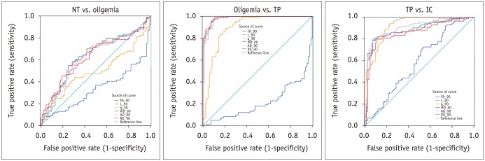
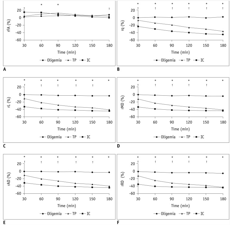
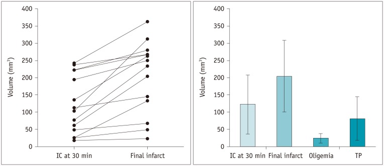
The mean volume of oligemia was 24.7 ± 14.1 mm, that of TP was 81.3 ± 62.6 mm, and that of IC was 123.0 ± 85.2 mm at 30 minutes after pMCAO. rFA showed an initial paradoxical 10% increase in IC and TP, and declined afterward. The rq, rL, rADC, rAD, and rRD showed an initial discrepant decrease in IC (from -24% to -36%) as compared with TP (from -7% to -13%). Significant differences ( < 0.05) in metrics, except rFA, were found between tissue subtypes in the first 2.5 hours. The rq demonstrated the best overall performance in discriminating TP from IC (accuracy = 92.6%, area under curve = 0.93) and the optimal cutoff value was -33.90%. The metric values for oligemia and NT remained similar at all time points.
Benign oligemia is small and remains microstructurally normal under pMCAO. TP and IC show a distinct evolution of DT-derived properties within the first 3 hours of stroke onset, and are thus potentially useful in predicting the fate of ischemic brain.
本研究旨在探讨在中风发作的前 3 小时内,良性低灌注、真正的“缺血半暗带”(TP)和梗死核心(IC)的扩散张量(DT)成像特征。
该研究得到了当地动物护理和使用委员会的批准。使用 Bruker 7T 磁共振扫描仪,在室气下对 14 只大鼠的永久性大脑中动脉闭塞(pMCAO)进行 DT 成像数据采集。每 30 分钟生成一次相对脑血流和表观扩散系数(ADC)图,以定义低灌注、TP、IC 和正常组织(NT)。通过与对侧正常大脑比较,得出相对各向异性分数(rFA)、纯各向异性(rq)、扩散幅度(rL)、ADC(rADC)、轴突扩散系数(rAD)和径向扩散系数(rRD)的值。
pMCAO 后 30 分钟时,低灌注区的平均体积为 24.7 ± 14.1mm,TP 区为 81.3 ± 62.6mm,IC 区为 123.0 ± 85.2mm。rFA 在 IC 和 TP 中最初出现了 10%的反常增加,之后则呈下降趋势。rq、rL、rADC、rAD 和 rRD 在 IC 中的初始差异下降幅度较大(从-24%到-36%),而在 TP 中则相对较小(从-7%到-13%)。在最初的 2.5 小时内,各组织类型之间的 rFA 以外的其他度量值存在显著差异(<0.05)。rq 在区分 TP 和 IC 方面表现最佳(准确性=92.6%,曲线下面积=0.93),最佳截断值为-33.90%。低灌注区和 NT 的测量值在所有时间点均保持相似。
pMCAO 下,良性低灌注体积较小,且组织结构仍正常。TP 和 IC 在中风发作的前 3 小时内具有明显不同的 DT 成像特征,因此可能有助于预测缺血性脑的预后。