Division of Neurology, Department of Medicine, University of British Columbia, Vancouver, British Columbia, Canada.
Department of Physics and Astronomy, University of British Columbia, Vancouver, British Columbia, Canada.
PLoS One. 2018 Nov 5;13(11):e0206607. doi: 10.1371/journal.pone.0206607. eCollection 2018.
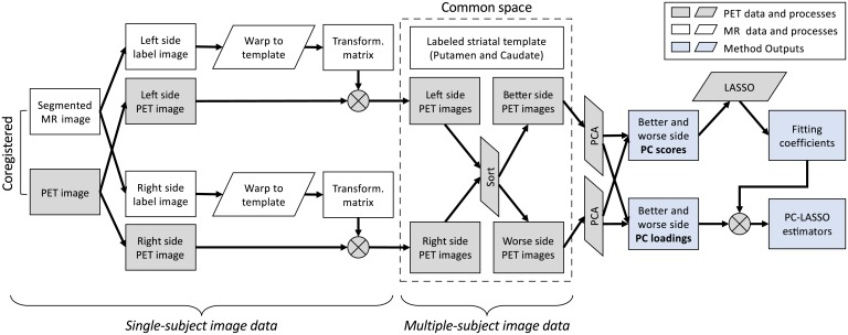
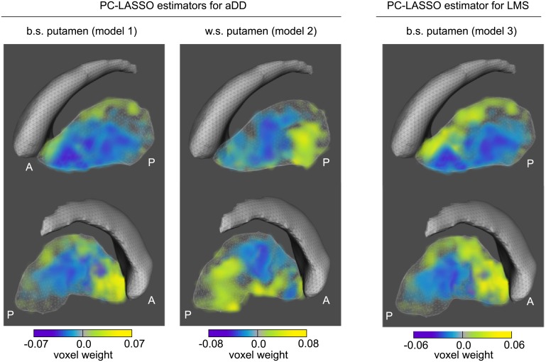
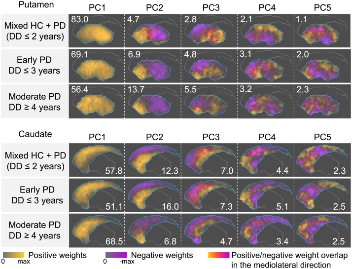
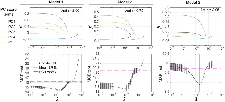
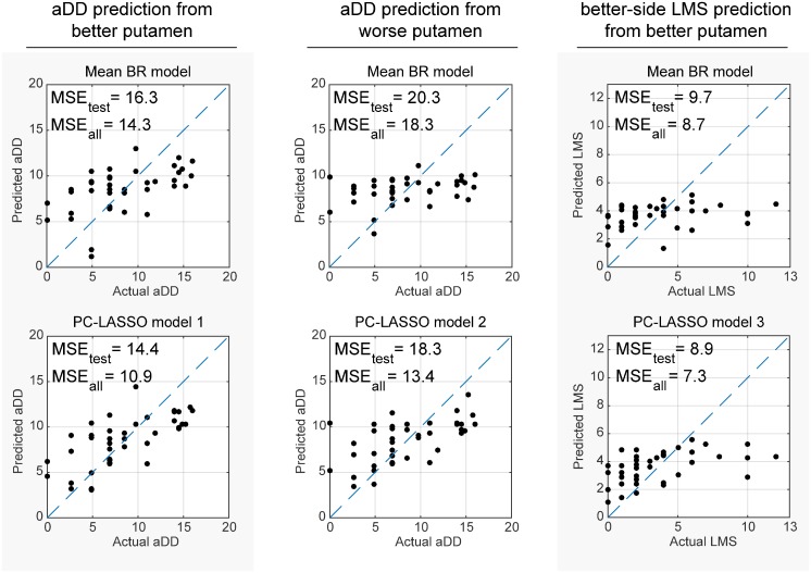
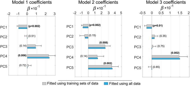
Spatial patterns of radiotracer binding in positron emission tomography (PET) images may convey information related to the disease topology. However, this information is not captured by the standard PET image analysis that quantifies the mean radiotracer uptake within a region of interest (ROI). On the other hand, spatial analyses that use more advanced radiomic features may be difficult to interpret. Here we propose an alternative data-driven, voxel-based approach to spatial pattern analysis in brain PET, which can be easily interpreted. We apply principal component analysis (PCA) to identify voxel covariance patterns, and optimally combine several patterns using the least absolute shrinkage and selection operator (LASSO). The resulting models predict clinical disease metrics from raw voxel values, allowing for inclusion of clinical covariates. The analysis is performed on high-resolution PET images from healthy controls and subjects affected by Parkinson's disease (PD), acquired with a pre-synaptic and a post-synaptic dopaminergic PET tracer. We demonstrate that PCA identifies robust and tracer-specific binding patterns in sub-cortical brain structures; the patterns evolve as a function of disease progression. Principal component LASSO (PC-LASSO) models of clinical disease metrics achieve higher predictive accuracy compared to the mean tracer binding ratio (BR) alone: the cross-validated test mean squared error of adjusted disease duration (motor impairment score) was 16.3 ± 0.17 years2 (9.7 ± 0.15) with mean BR, versus 14.4 ± 0.18 years2 (8.9 ± 0.16) with PC-LASSO. We interpret the best-performing PC-LASSO models in the spatial sense and discuss them with reference to the PD pathology and somatotopic organization of the striatum. PC-LASSO is thus shown to be a useful method to analyze clinically-relevant tracer binding patterns, and to construct interpretable, imaging-based predictive models of clinical metrics.
正电子发射断层扫描(PET)图像中示踪剂结合的空间模式可能传递与疾病拓扑结构相关的信息。然而,这一信息并未被标准的 PET 图像分析所捕捉,该分析仅量化了感兴趣区域(ROI)内的示踪剂平均摄取量。另一方面,使用更先进的放射组学特征的空间分析可能难以解释。在这里,我们提出了一种替代的、基于数据的、基于体素的方法来分析脑 PET 中的空间模式,这种方法易于解释。我们应用主成分分析(PCA)来识别体素协方差模式,并使用最小绝对值收缩和选择算子(LASSO)对几种模式进行最优组合。得到的模型从原始体素值预测临床疾病指标,允许纳入临床协变量。该分析是在健康对照者和帕金森病(PD)患者的高分辨率 PET 图像上进行的,这些图像是使用前突触和突触后多巴胺能 PET 示踪剂采集的。我们证明了 PCA 可以在皮质下脑结构中识别出稳健的、示踪剂特异性的结合模式;这些模式随着疾病的进展而演变。与单独使用示踪剂平均结合比(BR)相比,临床疾病指标的主成分 LASSO(PC-LASSO)模型达到了更高的预测准确性:经交叉验证的调整疾病持续时间(运动障碍评分)的测试均方误差为 16.3±0.17 年 2(9.7±0.15 年)与平均 BR 相比,而与 PC-LASSO 相比则为 14.4±0.18 年 2(8.9±0.16 年)。我们从空间意义上解释表现最好的 PC-LASSO 模型,并参考 PD 病理学和纹状体的躯体组织学讨论它们。因此,PC-LASSO 被证明是一种有用的方法,可以分析与临床相关的示踪剂结合模式,并构建基于成像的可解释的临床指标预测模型。