• 文献检索
  • 文档翻译
  • 深度研究
  • 学术资讯
  • Suppr Zotero 插件Zotero 插件
  • 邀请有礼
  • 套餐&价格
  • 历史记录
应用&插件
Suppr Zotero 插件Zotero 插件浏览器插件Mac 客户端Windows 客户端微信小程序
定价
高级版会员购买积分包购买API积分包
服务
文献检索文档翻译深度研究API 文档MCP 服务
关于我们
关于 Suppr公司介绍联系我们用户协议隐私条款
关注我们

Suppr 超能文献

核心技术专利:CN118964589B侵权必究
粤ICP备2023148730 号-1Suppr @ 2026

文献检索

告别复杂PubMed语法,用中文像聊天一样搜索,搜遍4000万医学文献。AI智能推荐,让科研检索更轻松。

立即免费搜索

文件翻译

保留排版,准确专业,支持PDF/Word/PPT等文件格式,支持 12+语言互译。

免费翻译文档

深度研究

AI帮你快速写综述,25分钟生成高质量综述,智能提取关键信息,辅助科研写作。

立即免费体验

混合PET/MR中的区域SUV定量:两种基于图谱的自动脑部分割方法的比较

Regional SUV quantification in hybrid PET/MR, a comparison of two atlas-based automatic brain segmentation methods.

作者信息

Ruan Weiwei, Sun Xun, Hu Xuehan, Liu Fang, Hu Fan, Guo Jinxia, Zhang Yongxue, Lan Xiaoli

机构信息

Department of Nuclear Medicine, Union Hospital, Tongji Medical College, Huazhong University of Science and Technology, No. 1277 Jiefang Ave, Wuhan, 430022, China.

Hubei Province Key Laboratory of Molecular Imaging, Wuhan, 430022, China.

出版信息

EJNMMI Res. 2020 Jun 8;10(1):60. doi: 10.1186/s13550-020-00648-8.

DOI:10.1186/s13550-020-00648-8
PMID:32514906
原文链接:https://pmc.ncbi.nlm.nih.gov/articles/PMC7280441/
Abstract

BACKGROUND

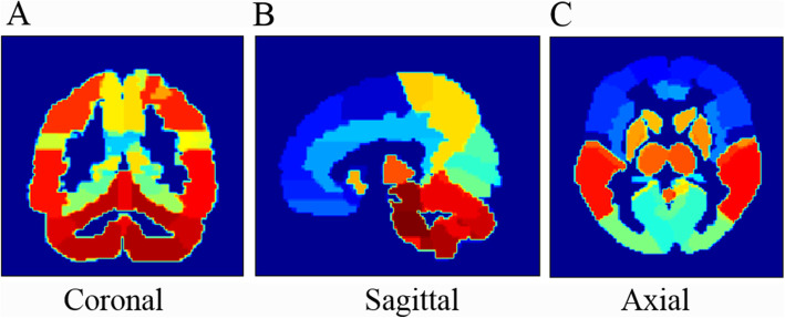
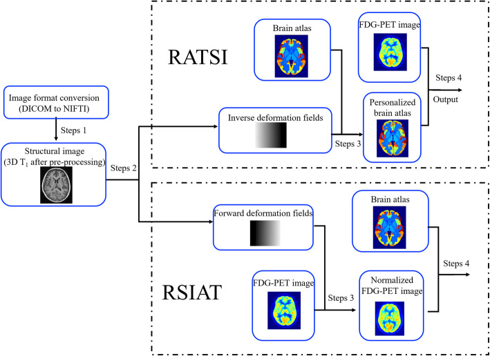
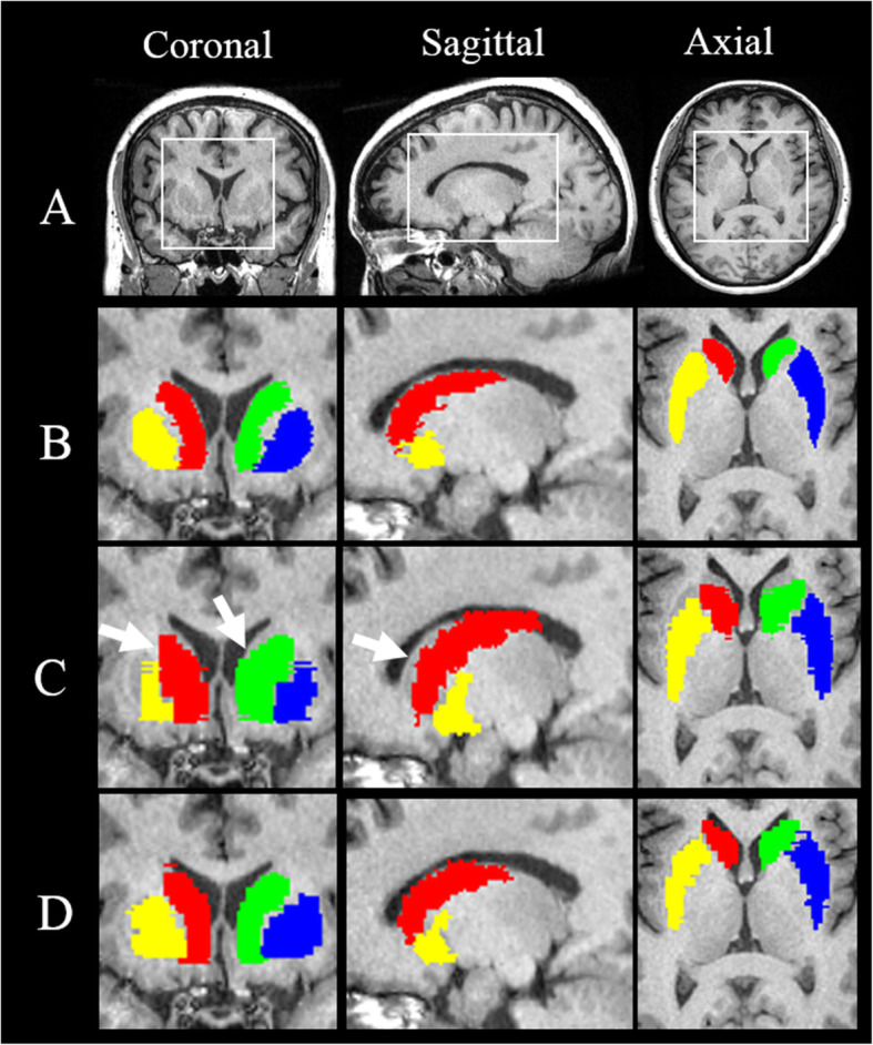
Quantitative analysis of brain positron-emission tomography (PET) depends on structural segmentation, which can be time-consuming and operator-dependent when performed manually. Previous automatic segmentation usually registered subjects' images onto an atlas template (defined as RSIAT here) for group analysis, which changed the individuals' images and probably affected regional PET segmentation. In contrast, we could register atlas template to subjects' images (RATSI), which created an individual atlas template and may be more accurate for PET segmentation. We segmented two representative brain areas in twenty Parkinson disease (PD) and eight multiple system atrophy (MSA) patients performed in hybrid positron-emission tomography/magnetic resonance imaging (PET/MR). The segmentation accuracy was evaluated using the Dice coefficient (DC) and Hausdorff distance (HD), and the standardized uptake value (SUV) measurements of these two automatic segmentation methods were compared, using manual segmentation as a reference.

RESULTS

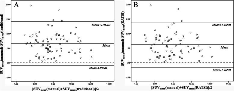
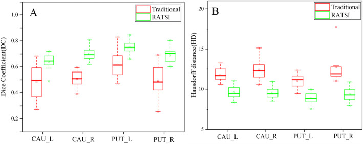
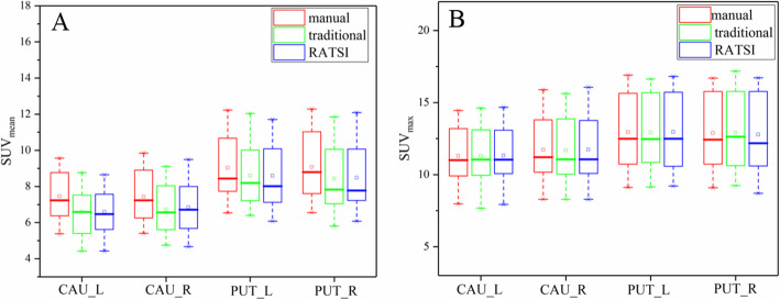
The DC of RATSI increased, and the HD decreased significantly (P < 0.05) compared with the RSIAT in PD, while the results of one-way analysis of variance (ANOVA) found no significant differences in the SUV and SUV among the two automatic and the manual segmentation methods. Further, RATSI was used to compare regional differences in cerebral metabolism pattern between PD and MSA patients. The SUV in the segmented cerebellar gray matter for the MSA group was significantly lower compared with the PD group (P < 0.05), which is consistent with previous reports.

CONCLUSION

The RATSI was more accurate for the caudate nucleus and putamen automatic segmentation and can be used for regional PET analysis in hybrid PET/MR.

摘要

背景

脑正电子发射断层扫描(PET)的定量分析依赖于结构分割,手动进行时既耗时又依赖操作人员。以往的自动分割通常将受试者的图像配准到一个图谱模板(此处定义为RSIAT)上进行组分析,这改变了个体图像,可能会影响PET区域分割。相比之下,我们可以将图谱模板配准到受试者的图像上(RATSI),从而创建个体图谱模板,这对于PET分割可能更准确。我们在20例帕金森病(PD)患者和8例多系统萎缩(MSA)患者中,对两个具有代表性的脑区进行了混合正电子发射断层扫描/磁共振成像(PET/MR)检查,并进行了分割。使用Dice系数(DC)和豪斯多夫距离(HD)评估分割准确性,并以手动分割为参考,比较这两种自动分割方法的标准化摄取值(SUV)测量结果。

结果

与PD患者的RSIAT相比,RATSI的DC增加,HD显著降低(P<0.05),而单因素方差分析(ANOVA)结果显示,两种自动分割方法和手动分割方法之间的SUV和SUV均无显著差异。此外,使用RATSI比较了PD和MSA患者脑代谢模式的区域差异。MSA组分割的小脑灰质SUV显著低于PD组(P<0.05),这与先前的报道一致。

结论

RATSI对尾状核和壳核的自动分割更准确,可用于混合PET/MR中的区域PET分析。

https://cdn.ncbi.nlm.nih.gov/pmc/blobs/e4dc/7280441/75f131b890d3/13550_2020_648_Fig6_HTML.jpg
https://cdn.ncbi.nlm.nih.gov/pmc/blobs/e4dc/7280441/d3b2a7cd3815/13550_2020_648_Fig1_HTML.jpg
https://cdn.ncbi.nlm.nih.gov/pmc/blobs/e4dc/7280441/695adf57300f/13550_2020_648_Fig2_HTML.jpg
https://cdn.ncbi.nlm.nih.gov/pmc/blobs/e4dc/7280441/2e0b3fb88847/13550_2020_648_Fig3_HTML.jpg
https://cdn.ncbi.nlm.nih.gov/pmc/blobs/e4dc/7280441/a977a889bdc3/13550_2020_648_Fig4_HTML.jpg
https://cdn.ncbi.nlm.nih.gov/pmc/blobs/e4dc/7280441/b50724a148a7/13550_2020_648_Fig5_HTML.jpg
https://cdn.ncbi.nlm.nih.gov/pmc/blobs/e4dc/7280441/75f131b890d3/13550_2020_648_Fig6_HTML.jpg

相似文献

1
Regional SUV quantification in hybrid PET/MR, a comparison of two atlas-based automatic brain segmentation methods.混合PET/MR中的区域SUV定量:两种基于图谱的自动脑部分割方法的比较
EJNMMI Res. 2020 Jun 8;10(1):60. doi: 10.1186/s13550-020-00648-8.
2
A multi-atlas based method for automated anatomical Macaca fascicularis brain MRI segmentation and PET kinetic extraction.基于多图谱的方法用于自动解剖猕猴大脑 MRI 分割和 PET 动力学提取。
Neuroimage. 2013 Aug 15;77:26-43. doi: 10.1016/j.neuroimage.2013.03.029. Epub 2013 Mar 26.
3
A multi-atlas based method for automated anatomical rat brain MRI segmentation and extraction of PET activity.一种基于多图谱的自动大鼠脑解剖MRI分割及PET活性提取方法。
PLoS One. 2014 Oct 17;9(10):e109113. doi: 10.1371/journal.pone.0109113. eCollection 2014.
4
Quantitative analysis of MRI-guided attenuation correction techniques in time-of-flight brain PET/MRI.基于磁共振成像的脑正电子发射断层扫描/磁共振成像中飞行时间技术的衰减校正的定量分析。
Neuroimage. 2016 Apr 15;130:123-133. doi: 10.1016/j.neuroimage.2016.01.060. Epub 2016 Feb 4.
5
Correction of quantification errors in pelvic and spinal lesions caused by ignoring higher photon attenuation of bone in [18F]NaF PET/MR.校正[18F]氟化钠正电子发射断层显像/磁共振成像中因忽视骨骼较高光子衰减而导致的盆腔和脊柱病变定量误差
Med Phys. 2015 Nov;42(11):6468-76. doi: 10.1118/1.4932367.
6
A computational pipeline for quantification of pulmonary infections in small animal models using serial PET-CT imaging.使用连续 PET-CT 成像对小动物模型中的肺部感染进行定量分析的计算流程。
EJNMMI Res. 2013 Jul 23;3(1):55. doi: 10.1186/2191-219X-3-55.
7
A Multi-Atlas-Based [18F]9-Fluoropropyl-(+)-Dihydrotetrabenazine Positron Emission Tomography Image Segmentation Method for Parkinson's Disease Quantification.一种基于多图谱的[18F]9-氟丙基-(+)-二氢丁苯那嗪正电子发射断层扫描图像分割方法用于帕金森病的量化分析。
Front Aging Neurosci. 2022 Jun 13;14:902169. doi: 10.3389/fnagi.2022.902169. eCollection 2022.
8
Impact of brain segmentation methods on regional metabolism quantification in F-FDG PET/MR analysis.脑部分割方法对F-FDG PET/MR分析中区域代谢定量的影响
EJNMMI Res. 2023 Sep 5;13(1):79. doi: 10.1186/s13550-023-01028-8.
9
Automatic Segmentation of Parkinson Disease Therapeutic Targets Using Nonlinear Registration and Clinical MR Imaging: Comparison of Methodology, Presence of Disease, and Quality Control.使用非线性配准和临床磁共振成像对帕金森病治疗靶点进行自动分割:方法学比较、疾病存在情况及质量控制
Stereotact Funct Neurosurg. 2023;101(2):146-157. doi: 10.1159/000526719. Epub 2023 Mar 7.
10
Multi-atlas active contour segmentation method using template optimization algorithm.基于模板优化算法的多图谱主动轮廓分割方法。
BMC Med Imaging. 2019 May 24;19(1):42. doi: 10.1186/s12880-019-0340-6.

引用本文的文献

1
Individualized diagnosis of Parkinson's disease based on multivariate magnetic resonance imaging radiomics and clinical indexes.基于多变量磁共振成像放射组学和临床指标的帕金森病个体化诊断
Front Aging Neurosci. 2025 Jan 31;17:1504733. doi: 10.3389/fnagi.2025.1504733. eCollection 2025.
2
Identification of Parkinson's disease and multiple system atrophy using multimodal PET/MRI radiomics.使用多模态 PET/MRI 放射组学识别帕金森病和多系统萎缩。
Eur Radiol. 2024 Jan;34(1):662-672. doi: 10.1007/s00330-023-10003-9. Epub 2023 Aug 3.
3
Deep learning segmentation results in precise delineation of the putamen in multiple system atrophy.

本文引用的文献

1
Metabolic pattern analysis of F-FDG PET as a marker for Parkinson's disease: a systematic review and meta-analysis.以¹⁸F-FDG PET代谢模式分析作为帕金森病标志物的系统评价与Meta分析
Rev Neurosci. 2019 May 3;30(7):743-756. doi: 10.1515/revneuro-2018-0061. Print 2019 Oct 25.
2
Quantitative Research of C-CFT and F-FDG PET in Parkinson's Disease: A Pilot Study With NeuroQ Software.帕金森病中C-CFT和F-FDG PET的定量研究:使用NeuroQ软件的初步研究
Front Neurosci. 2019 Apr 5;13:299. doi: 10.3389/fnins.2019.00299. eCollection 2019.
3
The diagnostic accuracy of F-FDG PET/CT in diagnosing fracture-related infections.
深度学习分割结果精确描绘了多系统萎缩中的壳核。
Eur Radiol. 2023 Oct;33(10):7160-7167. doi: 10.1007/s00330-023-09665-2. Epub 2023 May 1.
4
F-APN-1607 Tau Positron Emission Tomography Imaging for Evaluating Disease Progression in Alzheimer's Disease.F-APN-1607 Tau正电子发射断层成像用于评估阿尔茨海默病的疾病进展
Front Aging Neurosci. 2022 Feb 10;13:789054. doi: 10.3389/fnagi.2021.789054. eCollection 2021.
5
A Baboon Brain Atlas for Magnetic Resonance Imaging and Positron Emission Tomography Image Analysis.用于磁共振成像和正电子发射断层扫描图像分析的狒狒脑图谱。
Front Neuroanat. 2022 Jan 14;15:778769. doi: 10.3389/fnana.2021.778769. eCollection 2021.
18F-FDG PET/CT 诊断骨折相关感染的准确性。
Eur J Nucl Med Mol Imaging. 2019 Apr;46(4):999-1008. doi: 10.1007/s00259-018-4218-6. Epub 2018 Dec 7.
4
Data-driven, voxel-based analysis of brain PET images: Application of PCA and LASSO methods to visualize and quantify patterns of neurodegeneration.基于数据驱动的脑 PET 图像体素分析:PCA 和 LASSO 方法在可视化和量化神经退行性变模式中的应用。
PLoS One. 2018 Nov 5;13(11):e0206607. doi: 10.1371/journal.pone.0206607. eCollection 2018.
5
Optimization and comparative evaluation of nonlinear deformation algorithms for atlas-based segmentation of DBS target nuclei.基于图谱的 DBS 靶点分割的非线性变形算法的优化与比较评价。
Neuroimage. 2019 Jan 1;184:586-598. doi: 10.1016/j.neuroimage.2018.09.061. Epub 2018 Sep 26.
6
Classification of Alzheimer's and MCI Patients from Semantically Parcelled PET Images: A Comparison between AV45 and FDG-PET.基于语义分割PET图像的阿尔茨海默病和轻度认知障碍患者分类:AV45与FDG-PET的比较
Int J Biomed Imaging. 2018 Mar 15;2018:1247430. doi: 10.1155/2018/1247430. eCollection 2018.
7
Ultra High Field MRI-Guided Deep Brain Stimulation.超高场磁共振引导下的脑深部电刺激术
Trends Biotechnol. 2017 Oct;35(10):904-907. doi: 10.1016/j.tibtech.2017.06.010.
8
F-FDG PET in Parkinsonism: Differential Diagnosis and Evaluation of Cognitive Impairment.帕金森病中的氟代脱氧葡萄糖正电子发射断层显像:认知障碍的鉴别诊断与评估
J Nucl Med. 2017 Dec;58(12):1888-1898. doi: 10.2967/jnumed.116.186403. Epub 2017 Sep 14.
9
Toward defining deep brain stimulation targets in MNI space: A subcortical atlas based on multimodal MRI, histology and structural connectivity.朝向在 MNI 空间中定义深部脑刺激靶点:基于多模态 MRI、组织学和结构连接的皮质下图谱。
Neuroimage. 2018 Apr 15;170:271-282. doi: 10.1016/j.neuroimage.2017.05.015. Epub 2017 May 20.
10
Hybrid ZTE/Dixon MR-based attenuation correction for quantitative uptake estimation of pelvic lesions in PET/MRI.基于中兴通讯/狄克逊混合磁共振成像的PET/MRI盆腔病变定量摄取估计的衰减校正
Med Phys. 2017 Mar;44(3):902-913. doi: 10.1002/mp.12122.