• 文献检索
  • 文档翻译
  • 深度研究
  • 学术资讯
  • Suppr Zotero 插件Zotero 插件
  • 邀请有礼
  • 套餐&价格
  • 历史记录
应用&插件
Suppr Zotero 插件Zotero 插件浏览器插件Mac 客户端Windows 客户端微信小程序
定价
高级版会员购买积分包购买API积分包
服务
文献检索文档翻译深度研究API 文档MCP 服务
关于我们
关于 Suppr公司介绍联系我们用户协议隐私条款
关注我们

Suppr 超能文献

核心技术专利:CN118964589B侵权必究
粤ICP备2023148730 号-1Suppr @ 2026

文献检索

告别复杂PubMed语法,用中文像聊天一样搜索,搜遍4000万医学文献。AI智能推荐,让科研检索更轻松。

立即免费搜索

文件翻译

保留排版,准确专业,支持PDF/Word/PPT等文件格式,支持 12+语言互译。

免费翻译文档

深度研究

AI帮你快速写综述,25分钟生成高质量综述,智能提取关键信息,辅助科研写作。

立即免费体验

通过冷冻电子显微镜确定的细胞色素c亚硝酸还原酶的三维结构

Three-Dimensional Structure of Cytochrome c Nitrite Reductase As Determined by Cryo-Electron Microscopy.

作者信息

Baymukhametov T N, Chesnokov Y M, Pichkur E B, Boyko K M, Tikhonova T V, Myasnikov A G, Vasiliev A L, Lipkin A V, Popov V O, Kovalchuk M V

机构信息

National Research Center «Kurchatov Institute», Akademika Kurchatova Sqr., 1, Moscow, 123182 , Russia.

Bach Institute of Biochemistry, Research Center of Biotechnology of the Russian Academy of Sciences, Leninsky Ave., 33, bldg. 2, Moscow, 119071, Russia.

出版信息

Acta Naturae. 2018 Jul-Sep;10(3):48-56.

PMID:30397526
原文链接:https://pmc.ncbi.nlm.nih.gov/articles/PMC6209398/
Abstract

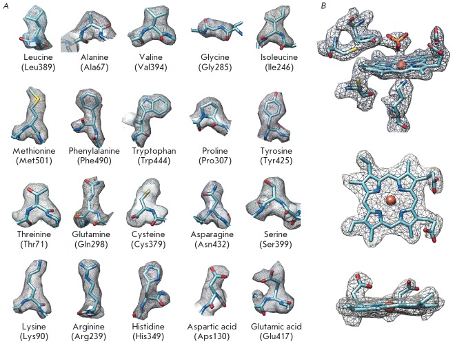
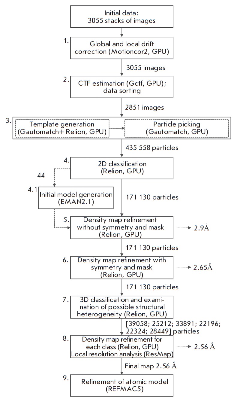
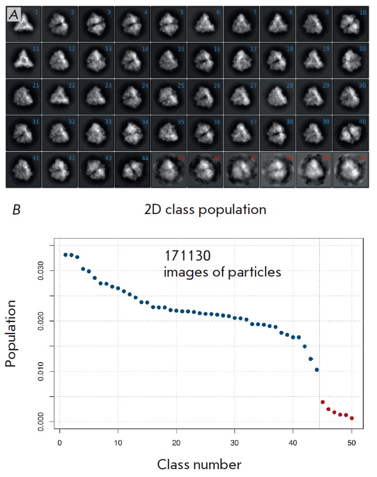
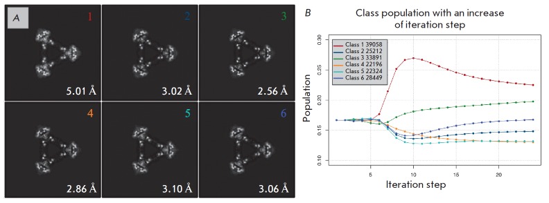
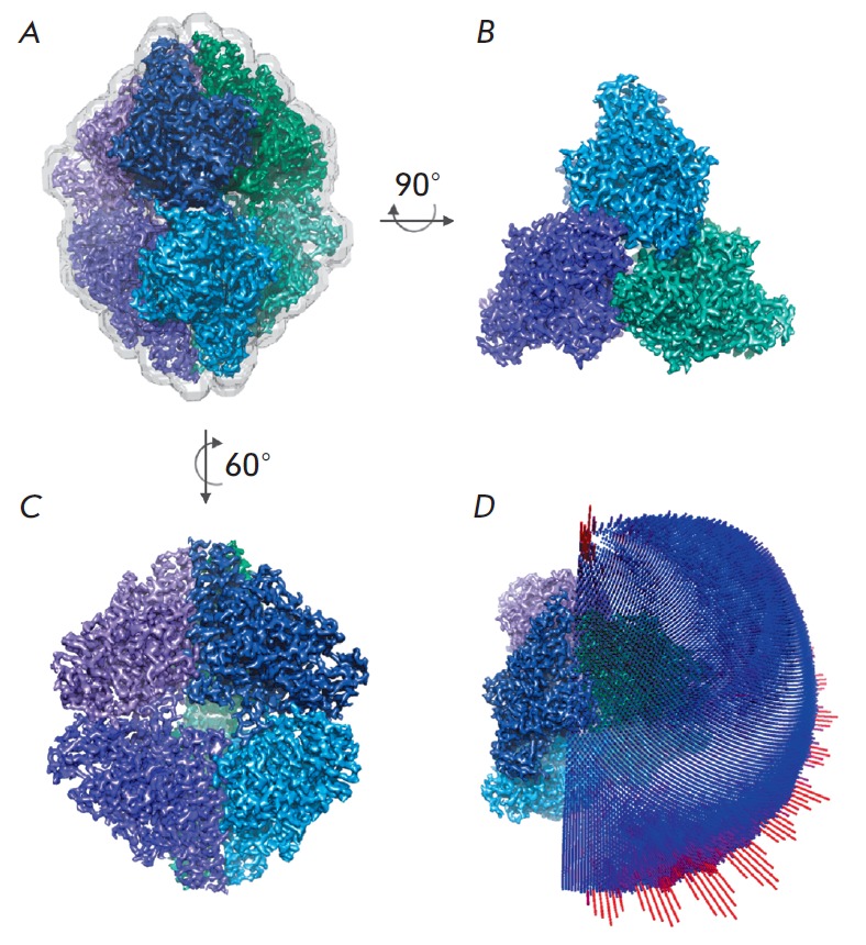
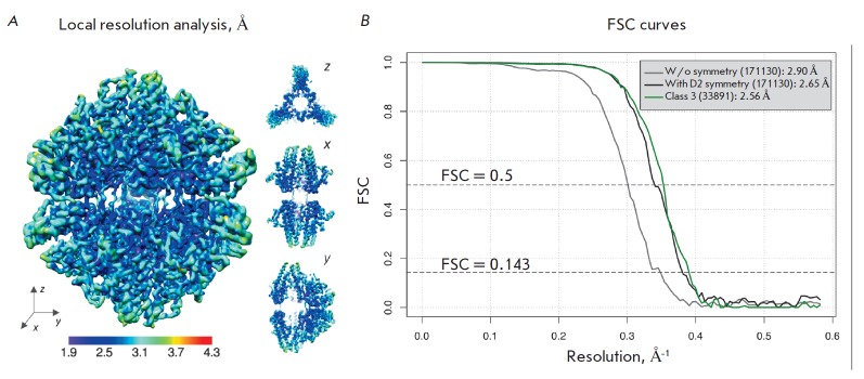
The structure of cytochrome c nitrite reductase from the bacterium Thioalkalivibrio nitratireducens was determined by cryo-electron microscopy (cryo-EM) at a 2.56 Å resolution. Possible structural heterogeneity of the enzyme was assessed. The backbone and side-chain orientations in the cryo-EM-based model are, in general, similar to those in the high-resolution X-ray diffraction structure of this enzyme.

摘要

通过冷冻电子显微镜(cryo-EM)在2.56埃分辨率下确定了来自嗜碱硫杆菌硝酸还原菌的细胞色素c亚硝酸盐还原酶的结构。评估了该酶可能存在的结构异质性。基于冷冻电子显微镜的模型中的主链和侧链取向通常与该酶的高分辨率X射线衍射结构中的取向相似。

https://cdn.ncbi.nlm.nih.gov/pmc/blobs/2493/6209398/25b956adaeae/AN20758251-10-3-048-g008.jpg
https://cdn.ncbi.nlm.nih.gov/pmc/blobs/2493/6209398/5d8bba2c9869/AN20758251-10-3-048-g001.jpg
https://cdn.ncbi.nlm.nih.gov/pmc/blobs/2493/6209398/92f1affca667/AN20758251-10-3-048-g002.jpg
https://cdn.ncbi.nlm.nih.gov/pmc/blobs/2493/6209398/92c86147fd68/AN20758251-10-3-048-g003.jpg
https://cdn.ncbi.nlm.nih.gov/pmc/blobs/2493/6209398/c3bda6890729/AN20758251-10-3-048-g004.jpg
https://cdn.ncbi.nlm.nih.gov/pmc/blobs/2493/6209398/a28b44ed2cca/AN20758251-10-3-048-g005.jpg
https://cdn.ncbi.nlm.nih.gov/pmc/blobs/2493/6209398/30146cad8d13/AN20758251-10-3-048-g006.jpg
https://cdn.ncbi.nlm.nih.gov/pmc/blobs/2493/6209398/a933ddbbde65/AN20758251-10-3-048-g007.jpg
https://cdn.ncbi.nlm.nih.gov/pmc/blobs/2493/6209398/25b956adaeae/AN20758251-10-3-048-g008.jpg

相似文献

1
Three-Dimensional Structure of Cytochrome c Nitrite Reductase As Determined by Cryo-Electron Microscopy.通过冷冻电子显微镜确定的细胞色素c亚硝酸还原酶的三维结构
Acta Naturae. 2018 Jul-Sep;10(3):48-56.
2
High-resolution structural analysis of a novel octaheme cytochrome c nitrite reductase from the haloalkaliphilic bacterium Thioalkalivibrio nitratireducens.来自嗜盐碱细菌嗜硫代碱弧菌的新型八血红素细胞色素c亚硝酸盐还原酶的高分辨率结构分析。
J Mol Biol. 2009 Jun 26;389(5):846-62. doi: 10.1016/j.jmb.2009.04.037. Epub 2009 Apr 23.
3
Isolation and oligomeric composition of cytochrome c nitrite reductase from the haloalkaliphilic bacterium Thioalkalivibrio nitratireducens.从嗜盐碱细菌硫代碱弧菌硝酸盐还原菌中分离细胞色素c亚硝酸还原酶及其寡聚体组成
Biochemistry (Mosc). 2008 Feb;73(2):164-70. doi: 10.1134/s0006297908020077.
4
2.85 and 2.99 Å resolution structures of 110 kDa nitrite reductase determined by 200 kV cryogenic electron microscopy.200kV 低温电子显微镜测定的 110 kDa 亚硝酸盐还原酶的 2.85 和 2.99Å分辨率结构。
J Struct Biol. 2021 Sep;213(3):107768. doi: 10.1016/j.jsb.2021.107768. Epub 2021 Jul 1.
5
Structural study of the X-ray-induced enzymatic reaction of octahaem cytochrome C nitrite reductase.八聚体细胞色素C亚硝酸还原酶X射线诱导酶促反应的结构研究
Acta Crystallogr D Biol Crystallogr. 2015 May;71(Pt 5):1087-94. doi: 10.1107/S1399004715003053. Epub 2015 Apr 24.
6
Structures of complexes of octahaem cytochrome c nitrite reductase from Thioalkalivibrio nitratireducens with sulfite and cyanide.
Acta Crystallogr D Biol Crystallogr. 2010 Oct;66(Pt 10):1043-7. doi: 10.1107/S0907444910031665. Epub 2010 Sep 18.
7
Rapid increase of near atomic resolution virus capsid structures determined by cryo-electron microscopy.通过冷冻电镜快速解析近原子分辨率的病毒衣壳结构。
J Struct Biol. 2018 Jan;201(1):1-4. doi: 10.1016/j.jsb.2017.10.007. Epub 2017 Oct 27.
8
High-Resolution Single-Molecule Structure Revealed by Electron Microscopy and Individual Particle Electron Tomography.通过电子显微镜和单颗粒电子断层扫描揭示的高分辨率单分子结构
J Phys Chem Biophys. 2012;2(2). doi: 10.4172/2161-0398.1000e103. Epub 2012 May 22.
9
Determining the Crystal Structure of TRPV6确定瞬时受体电位香草酸亚型6(TRPV6)的晶体结构
10
High-resolution structure of phosphoketolase from Bifidobacterium longum determined by cryo-EM single-particle analysis.通过 cryo-EM 单颗粒分析确定长双歧杆菌磷酸酮醇酶的高分辨率结构。
J Struct Biol. 2022 Jun;214(2):107842. doi: 10.1016/j.jsb.2022.107842. Epub 2022 Feb 15.

引用本文的文献

1
11168H Exposed to Penicillin Forms Persister Cells and Cells With Altered Redox Protein Activity.11168H 接触青霉素后形成持留细胞和氧化还原蛋白活性改变的细胞。
Front Cell Infect Microbiol. 2020 Oct 19;10:565975. doi: 10.3389/fcimb.2020.565975. eCollection 2020.

本文引用的文献

1
MonoRes: Automatic and Accurate Estimation of Local Resolution for Electron Microscopy Maps.MonoRes:自动且精确估计电子显微镜图谱的局部分辨率。
Structure. 2018 Feb 6;26(2):337-344.e4. doi: 10.1016/j.str.2017.12.018. Epub 2018 Jan 27.
2
Cryo-electron microscopy structure of the lysosomal calcium-permeable channel TRPML3.溶酶体钙通透通道TRPML3的冷冻电镜结构
Nature. 2017 Oct 19;550(7676):411-414. doi: 10.1038/nature24055. Epub 2017 Oct 11.
3
Accelerated cryo-EM structure determination with parallelisation using GPUs in RELION-2.
在RELION-2中使用图形处理器(GPU)并行化加速冷冻电镜结构测定
Elife. 2016 Nov 15;5:e18722. doi: 10.7554/eLife.18722.
4
Single-Particle Refinement and Variability Analysis in EMAN2.1.EMAN2.1中的单颗粒精修与变异性分析
Methods Enzymol. 2016;579:159-89. doi: 10.1016/bs.mie.2016.05.001. Epub 2016 Jul 1.
5
Processing of Structurally Heterogeneous Cryo-EM Data in RELION.RELION中结构异质冷冻电镜数据的处理
Methods Enzymol. 2016;579:125-57. doi: 10.1016/bs.mie.2016.04.012. Epub 2016 May 31.
6
Breaking Cryo-EM Resolution Barriers to Facilitate Drug Discovery.突破冷冻电镜分辨率障碍以促进药物发现。
Cell. 2016 Jun 16;165(7):1698-1707. doi: 10.1016/j.cell.2016.05.040. Epub 2016 May 26.
7
Gctf: Real-time CTF determination and correction.Gctf:实时对比度传递函数(CTF)测定与校正
J Struct Biol. 2016 Jan;193(1):1-12. doi: 10.1016/j.jsb.2015.11.003. Epub 2015 Nov 19.
8
The revolution will not be crystallized: a new method sweeps through structural biology.这场革命不会被固化:一种新方法席卷结构生物学领域。
Nature. 2015 Sep 10;525(7568):172-4. doi: 10.1038/525172a.
9
Measuring the optimal exposure for single particle cryo-EM using a 2.6 Å reconstruction of rotavirus VP6.利用轮状病毒VP6的2.6埃分辨率重建来测量单颗粒冷冻电镜的最佳曝光量。
Elife. 2015 May 29;4:e06980. doi: 10.7554/eLife.06980.
10
Single-Particle Cryo-EM at Crystallographic Resolution.达到晶体学分辨率的单颗粒冷冻电镜技术。
Cell. 2015 Apr 23;161(3):450-457. doi: 10.1016/j.cell.2015.03.049.