Department of Biomedical Sciences, Institute of Radiological Sciences, University of Sassari, Sassari, Italy.
Department of Clinical and Experimental Medicine, Institute of Radiological Sciences, University of Sassari, Sassari, Italy.
Biomed Res Int. 2018 Oct 8;2018:6120703. doi: 10.1155/2018/6120703. eCollection 2018.
Over the last decade, the field of medical imaging experienced an exponential growth, leading to the development of radiomics, with which innumerable quantitative features are obtained from digital medical images, providing a comprehensive characterization of the tumor. This review aims to assess the role of this emerging diagnostic tool in breast cancer, focusing on the ability of radiomics to predict malignancy, response to neoadjuvant chemotherapy, prognostic factors, molecular subtypes, and risk of recurrence.
A literature search on PubMed and on Cochrane database websites to retrieve English-written systematic reviews, review articles, meta-analyses, and randomized clinical trials published from August 2013 up to July 2018 was carried out.
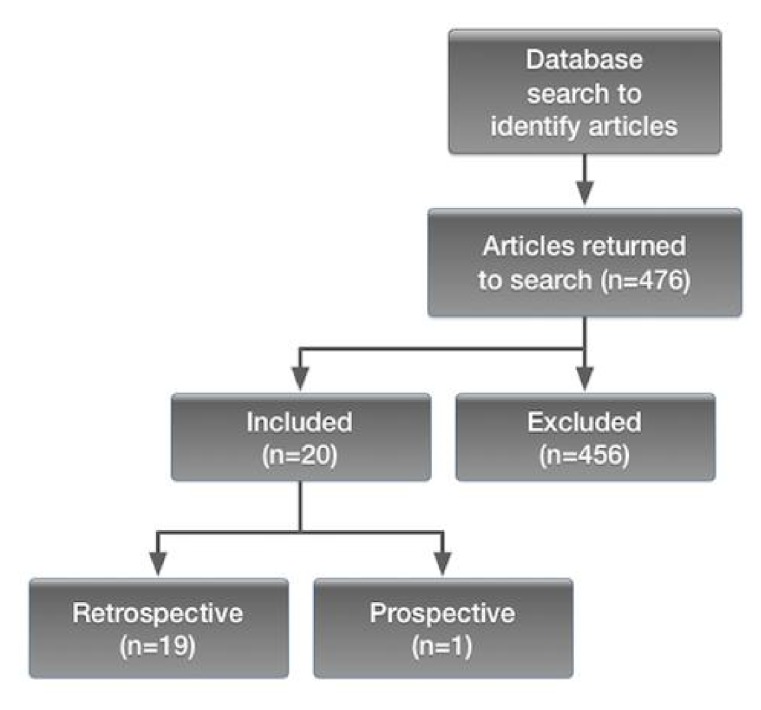
Twenty papers (19 retrospective and 1 prospective studies) conducted with different conventional imaging modalities were included.
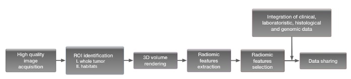
The integration of quantitative information with clinical, histological, and genomic data could enable clinicians to provide personalized treatments for breast cancer patients. Current limitations of a routinely application of radiomics are represented by the limited knowledge of its basics concepts among radiologists and by the lack of efficient and standardized systems of feature extraction and data sharing.
过去十年中,医学影像学领域经历了指数级的增长,由此产生了放射组学,它可以从数字医学图像中获得无数的定量特征,对肿瘤进行全面的描述。本综述旨在评估这一新兴诊断工具在乳腺癌中的作用,重点关注放射组学在预测恶性肿瘤、新辅助化疗反应、预后因素、分子亚型和复发风险方面的能力。
在 PubMed 和 Cochrane 数据库网站上进行了文献检索,以检索 2013 年 8 月至 2018 年 7 月期间发表的英文系统评价、综述文章、荟萃分析和随机临床试验。
纳入了 20 篇论文(19 项回顾性研究和 1 项前瞻性研究),这些研究使用了不同的常规成像方式。
将定量信息与临床、组织学和基因组数据相结合,可以使临床医生为乳腺癌患者提供个性化的治疗方案。目前,放射组学的常规应用受到一些限制,包括放射科医生对其基本概念的了解有限,以及缺乏高效和标准化的特征提取和数据共享系统。