Ye Ling, Wen Chuanbing, Liu Hui
Department of Pain management, West China Hospital, Sichuan University, Chengdu, Sichuan Province, 610041, People's Republic of China.
Department of Pain Management, Sichuan Academy of Medical Sciences & Sichuan Provincial People's Hospital, Chengdu, Sichuan Province, 610072, People's Republic of China.
BMC Anesthesiol. 2018 Nov 7;18(1):160. doi: 10.1186/s12871-018-0620-7.
The purpose of this study was to investigate the feasibility, accuracy and efficiency of the facet joint injections in the lumbar spine by ultrasound guided versus lose dose computed tomography (CT) guidance.
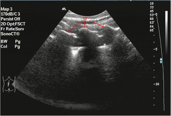
First the examination on the joint space of the facet joints of the lumbar spine was obtained by the ultrasound in 10 patients. Second forty patients were randomized assigned into two groups: ultrasound group and low dose CT group. Comparison was made in the clinical efficiency between the ultrasound-guided group and CT group. The feasibility, accuracy and efficiency of the ultrasound-guided lumbar facet joint injections were also evaluated.
A total of 88 lumbar facet joints from L to S were clearly visualized in the 10 patients. Both the ultrasound and the CT measurements showed the same average depth and lateral distance to the reference point (P > 0.05). And 86.5% of the facet joint injections (64/74) were correctly performed under the ultrasound guidance in the first time. The exact placement of the needle tips was evaluated by CT. After the lumbar facet joint injections, the clinical efficiency was almost the same in the ultrasound-guided group as in the CT group.
The lumbar facet joint space can be accurately demonstrated by ultrasound. The ultrasound-guided facet joint injection in the lumbar spine obtained almost the same satisfactory feasibility, accuracy and clinical efficiency compared with low dose CT. Ultrasound technique could provide the real-time monitoring.
This study was registered on Chinese Clinical Trial Registry ( ChiCTR1800018819 , retrospective registered on 11/10/2018).
本研究旨在探讨超声引导与低剂量计算机断层扫描(CT)引导下腰椎小关节注射的可行性、准确性和效率。
首先对10例患者进行超声检查以获取腰椎小关节间隙情况。其次将40例患者随机分为两组:超声组和低剂量CT组。比较超声引导组和CT组的临床效果。同时评估超声引导下腰椎小关节注射的可行性、准确性和效率。
10例患者中,从L到S的88个腰椎小关节均清晰可见。超声和CT测量显示到参考点的平均深度和横向距离相同(P>0.05)。在超声引导下,首次进行小关节注射时,86.5%(64/74)操作正确。通过CT评估针尖的准确位置。腰椎小关节注射后,超声引导组与CT组的临床效果几乎相同。
超声可准确显示腰椎小关节间隙。与低剂量CT相比,超声引导下腰椎小关节注射在可行性、准确性和临床效果方面几乎同样令人满意。超声技术可提供实时监测。
本研究在中国临床试验注册中心注册(ChiCTR1800018819,2018年10月11日回顾性注册)。