Key Laboratory of Animal Epidemiology of the Ministry of Agriculture, College of Veterinary Medicine and State Key Laboratory of Agrobiotechnology, China Agricultural University, No.2 Yuanmingyuan West Road, Haidian Distract, Beijing, 100193, People's Republic of China.
Animal Medicine Research Center of DBN Group, South Crossroad of Xiangrui Street and Huatuo Road DBN Daxing Science Park, Daxing Distract, Beijing, 102600, People's Republic of China.
Virol J. 2018 Nov 7;15(1):170. doi: 10.1186/s12985-018-1078-4.
Porcine Epidemic Diarrhea (PED) is an acute and highly contagious enteric disease caused by PED virus (PEDV), characterized by vomitting, watery diarrhea and fatal dehydration with high mortality in sucking piglets of one week of age. Although PEDV induced cell apoptosis has been established in vitro and in vivo, the functional protein that contributes to this event remains unclear.
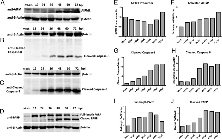
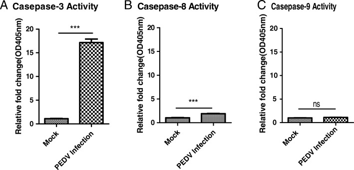
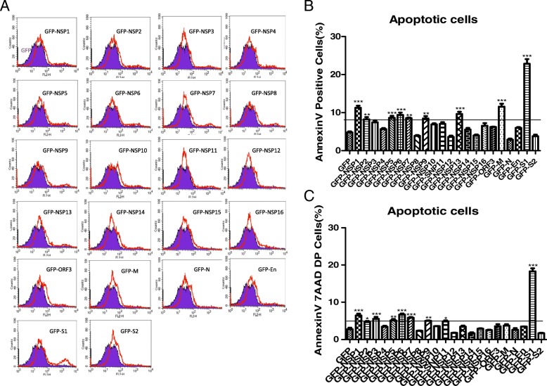
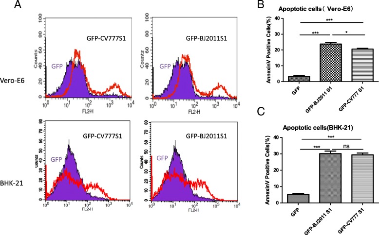
The activation or cleavage of main apoptosis-associated molecular such as AIFM1, caspase-3, caspase-8, caspase-9 and PARP in PEDV infected host cells were analyzed by western blotting. The nuclear change of infected cell was monitored by confocal immunofluorescence assay. The overexpressing plasmids of 16 non-structural proteins (Nsp1-16) and 6 structural proteins (M, N, E, ORF3, S1 and S2) were constructed by cloning. Cell apoptosis induced by PEDV or overexpression non-structural or structural proteins was measured by the flow cytometry assay.
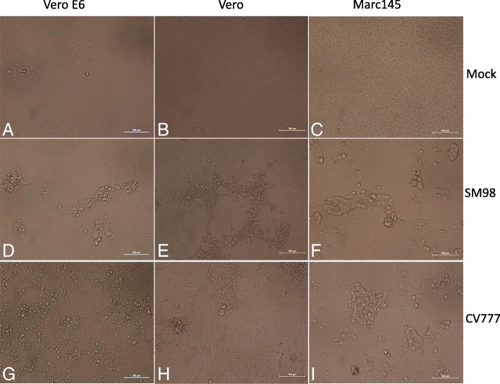
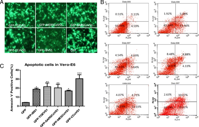
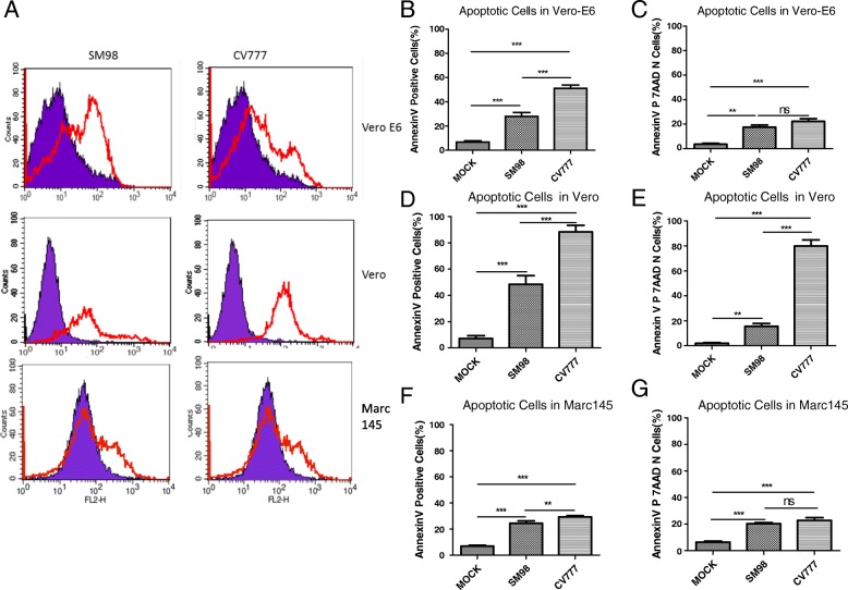
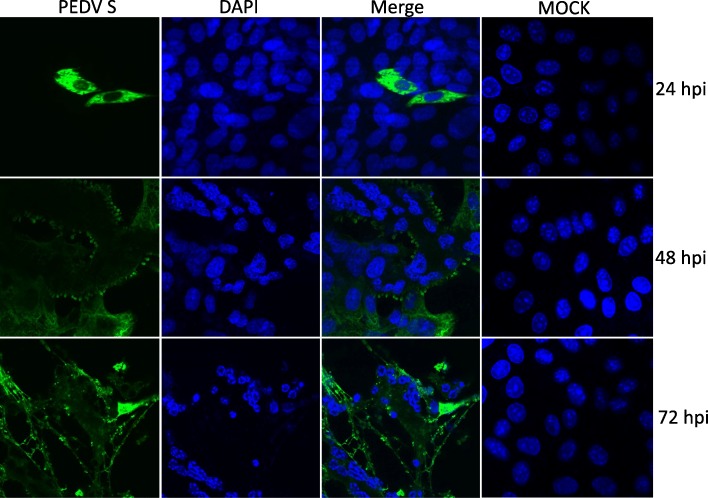
PEDV could infect various host cells including Vero, Vero-E6 and Marc-145 and cause obvious cytopathic effects, including roundup, cell fusion, cell membrane vacuolation, syncytium formation and cause apparent apoptosis. In infected cells, PEDV-induced apoptosis is accompanied by nuclear concentration and fragmentation as a result of caspase-3 and caspase-8 activation and AIFM1 and PARP cleavage. Overexpression of S1 Spike protein of PEDV SM98 strain effectively induced host cell apoptosis, while the expression of the other non-structure proteins (Nsp1-16) and structural proteins (M, N, E, S2 and ORF3) has no or less effect on cell apoptosis. Similarly, expression of S1 protein from wild-type strain BJ2011 or cell-adapted strain CV777, also induce apoptosis in transfected cells. Finally, we demonstrated that the S1 proteins from various coronavirus family members such as TGEV, IBV, CCoV, SARS and MERS could also induce Vero-E6 cells apoptosis.
S1 Spike protein is one of the most critical functional proteins that contribute to cell apoptosis. Expression of S1 proteins of the coronavirus tested in this study could all induce cell apoptosis suggesting S1 maybe is an effective inducer in Coronavirus-induced cell apoptosis and targeting S1 protein expression probably is a promising strategy to inhibit coronavirus infection and thus mediated apoptosis on host cells.
猪流行性腹泻(PED)是由猪流行性腹泻病毒(PEDV)引起的一种急性、高度传染性的肠病,以呕吐、水样腹泻和 1 周龄仔猪致命性脱水为特征,死亡率很高。虽然 PEDV 诱导的细胞凋亡已在体外和体内得到证实,但导致这种现象的功能蛋白尚不清楚。
通过 Western blot 分析了 PEDV 感染宿主细胞中主要凋亡相关分子如 AIFM1、caspase-3、caspase-8、caspase-9 和 PARP 的激活或切割。通过共聚焦免疫荧光检测感染细胞的核变化。通过克隆构建了 16 种非结构蛋白(Nsp1-16)和 6 种结构蛋白(M、N、E、ORF3、S1 和 S2)的过表达质粒。通过流式细胞术检测 PEDV 或过表达非结构或结构蛋白诱导的细胞凋亡。
PEDV 可感染多种宿主细胞,包括 Vero、Vero-E6 和 Marc-145,并引起明显的细胞病变效应,包括细胞圆化、细胞融合、细胞膜空泡化、合胞体形成,并导致明显的细胞凋亡。在感染细胞中,PEDV 诱导的凋亡伴随着 caspase-3 和 caspase-8 的激活以及 AIFM1 和 PARP 的切割导致核浓缩和碎片化。PEDV SM98 株 S1 刺突蛋白的过表达有效地诱导了宿主细胞凋亡,而其他非结构蛋白(Nsp1-16)和结构蛋白(M、N、E、S2 和 ORF3)的表达对细胞凋亡没有或影响较小。同样,野生型 BJ2011 株或细胞适应株 CV777 的 S1 蛋白的表达也可诱导转染细胞凋亡。最后,我们证明了来自 TGEV、IBV、CCoV、SARS 和 MERS 等冠状病毒家族成员的 S1 蛋白也可诱导 Vero-E6 细胞凋亡。
S1 刺突蛋白是导致细胞凋亡的关键功能蛋白之一。本研究中检测的冠状病毒的 S1 蛋白表达均可诱导细胞凋亡,提示 S1 可能是冠状病毒诱导细胞凋亡的有效诱导剂,靶向 S1 蛋白表达可能是抑制冠状病毒感染和介导宿主细胞凋亡的有前途的策略。