Verloh Niklas, Einspieler Ingo, Utpatel Kirsten, Menhart Karin, Brunner Stefan, Hofheinz Frank, van den Hoff Jörg, Wiggermann Philipp, Evert Matthias, Stroszczynski Christian, Hellwig Dirk, Grosse Jirka
Department of Nuclear Medicine, University Hospital Regensburg, Regensburg, Germany.
Department of Radiology, University Hospital Regensburg, Regensburg, Germany.
EJNMMI Res. 2018 Nov 9;8(1):98. doi: 10.1186/s13550-018-0452-y.
The aim of this study was to assess the value of F-FDG PET/CT for quantitative assessment of hepatic metabolism in patients with different stages of liver fibrosis/cirrhosis.
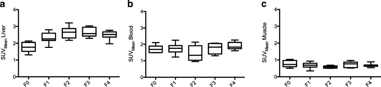
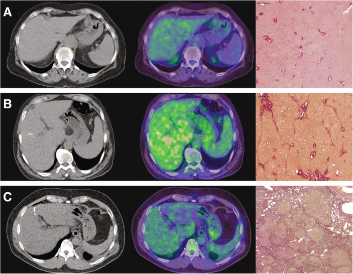
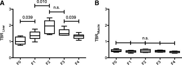
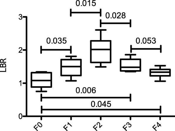
F-FDG PET/CT scans of 37 patients either with or without liver fibrosis/cirrhosis, classified according to the METAVIR score (F0-F4) obtained from histopathological analysis of liver specimen, were analyzed retrospectively and classified as follows: no liver fibrosis (F0, n = 6), mild liver fibrosis (F1, n = 11), advanced liver fibrosis (F2, n = 6), severe liver fibrosis (F3, n = 5), and liver cirrhosis (F4, n = 11). The liver-to-blood ratio (LBR, scan time corrected for a reference time of 75 min) was compared between patient groups.
Patients with liver fibrosis or cirrhosis (≥ F1; LBR 1.53 ± 0.35) showed a significant higher LBR than patients with normal liver parenchyma (F0, 1.08 ± 0.23; P = 0.004). In direct comparison, LBR increased up to the advanced stage of liver fibrosis (F2; 2.00 ± 0.40) and decreased until liver cirrhosis is reached (F4, 1.32 ± 0.14).
Functional changes in liver parenchyma during liver fibrosis/cirrhosis affect hepatic glucose metabolism and significantly differ between stages of liver fibrosis/cirrhosis, classified according to the METAVIR scoring system, as demonstrated by LBR quantification by F-FDG PET/CT.
本研究旨在评估F-FDG PET/CT对不同阶段肝纤维化/肝硬化患者肝脏代谢进行定量评估的价值。
回顾性分析37例有或无肝纤维化/肝硬化患者的F-FDG PET/CT扫描结果,根据肝脏标本组织病理学分析获得的METAVIR评分(F0-F4)进行分类,具体如下:无肝纤维化(F0,n = 6)、轻度肝纤维化(F1,n = 11)、进展期肝纤维化(F2,n = 6)、重度肝纤维化(F3,n = 5)和肝硬化(F4,n = 11)。比较各患者组之间的肝血比(LBR,扫描时间校正为参考时间75分钟)。
肝纤维化或肝硬化患者(≥F1;LBR 1.53±0.35)的LBR显著高于肝实质正常的患者(F0,1.08±0.23;P = 0.004)。直接比较发现,LBR在进展期肝纤维化(F2;2.00±0.40)时升高,直至肝硬化(F4,1.32±0.14)时降低。
F-FDG PET/CT通过LBR定量显示,肝纤维化/肝硬化过程中肝实质的功能变化会影响肝脏葡萄糖代谢,且在根据METAVIR评分系统分类的肝纤维化/肝硬化各阶段之间存在显著差异。