University of Lincoln, School of Computer Science, Lincoln, UK.
University of Lincoln, School of Computer Science, Lincoln, UK.
Phys Med. 2018 Nov;55:149-154. doi: 10.1016/j.ejmp.2018.10.020. Epub 2018 Nov 9.
Proton CT is widely recognised as a beneficial alternative to conventional X-ray CT for treatment planning in proton beam radiotherapy. A novel proton CT imaging system, based entirely on solid-state detector technology, is presented. Compared to conventional scintillator-based calorimeters, positional sensitive detectors allow for multiple protons to be tracked per read out cycle, leading to a potential reduction in proton CT scan time. Design and characterisation of its components are discussed. An early proton CT image obtained with a fully solid-state imaging system is shown and accuracy (as defined in Section IV) in Relative Stopping Power to water (RSP) quantified.
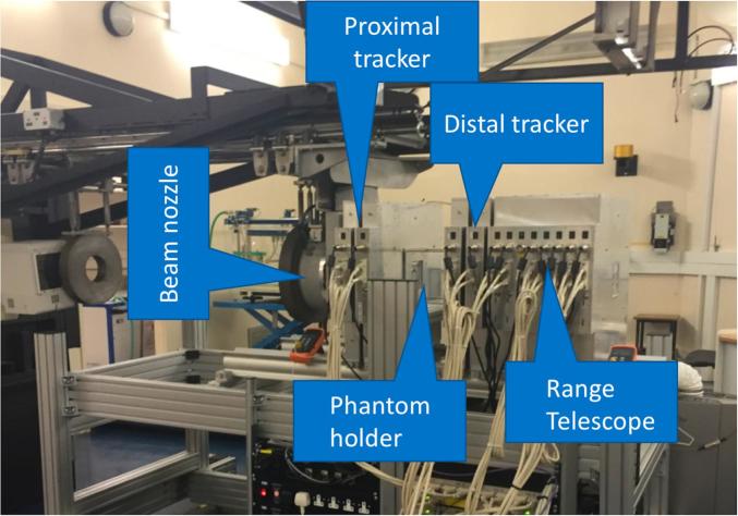
A solid-state imaging system for proton CT, based on silicon strip detectors, has been developed by the PRaVDA collaboration. The system comprises a tracking system that infers individual proton trajectories through an imaging phantom, and a Range Telescope (RT) which records the corresponding residual energy (range) for each proton. A back-projection-then-filtering algorithm is used for CT reconstruction of an experimentally acquired proton CT scan.
An initial experimental result for proton CT imaging with a fully solid-state system is shown for an imaging phantom, namely a 75 mm diameter PMMA sphere containing tissue substitute inserts, imaged with a passively-scattered 125 MeV beam. Accuracy in RSP is measured to be ⩽1.6% for all the inserts shown.
A fully solid-state imaging system for proton CT has been shown capable of imaging a phantom with protons and successfully improving RSP accuracy. These promising results, together with system the capability to cope with high proton fluences (2×10 protons/s), suggests that this research platform could improve current standards in treatment planning for proton beam radiotherapy.
质子 CT 被广泛认为是质子束放射治疗中治疗计划的一种有益替代方案,优于传统的 X 射线 CT。本文提出了一种完全基于固态探测器技术的新型质子 CT 成像系统。与传统的闪烁体基量热计相比,位置灵敏探测器允许每个读出周期跟踪多个质子,从而有可能减少质子 CT 扫描时间。讨论了其组件的设计和特性。展示了使用完全固态成像系统获得的早期质子 CT 图像,并量化了水中相对阻止本领(RSP)的准确性(如第 IV 部分所述)。
PRaVDA 合作开发了一种基于硅条探测器的质子 CT 固态成像系统。该系统包括一个通过成像体模推断单个质子轨迹的跟踪系统,以及一个记录每个质子相应剩余能量(射程)的射程望远镜(RT)。使用反向投影然后滤波算法对实验获得的质子 CT 扫描进行 CT 重建。
展示了一个完全固态系统的质子 CT 成像的初始实验结果,该系统对一个成像体模进行成像,即一个包含组织替代物插入物的 75mm 直径 PMMA 球体,使用被动散射的 125MeV 束进行成像。对于所有显示的插入物,RSP 的准确性测量值 ⩽1.6%。
已经证明,一种完全固态的质子 CT 成像系统能够对质子进行成像,并成功提高了 RSP 准确性。这些有希望的结果,以及系统能够应对高质子通量(2×10 个质子/s)的能力,表明该研究平台可以提高质子束放射治疗中当前的治疗计划标准。