• 文献检索
  • 文档翻译
  • 深度研究
  • 学术资讯
  • Suppr Zotero 插件Zotero 插件
  • 邀请有礼
  • 套餐&价格
  • 历史记录
应用&插件
Suppr Zotero 插件Zotero 插件浏览器插件Mac 客户端Windows 客户端微信小程序
定价
高级版会员购买积分包购买API积分包
服务
文献检索文档翻译深度研究API 文档MCP 服务
关于我们
关于 Suppr公司介绍联系我们用户协议隐私条款
关注我们

Suppr 超能文献

核心技术专利:CN118964589B侵权必究
粤ICP备2023148730 号-1Suppr @ 2026

文献检索

告别复杂PubMed语法,用中文像聊天一样搜索,搜遍4000万医学文献。AI智能推荐,让科研检索更轻松。

立即免费搜索

文件翻译

保留排版,准确专业,支持PDF/Word/PPT等文件格式,支持 12+语言互译。

免费翻译文档

深度研究

AI帮你快速写综述,25分钟生成高质量综述,智能提取关键信息,辅助科研写作。

立即免费体验

肾管状上皮细胞:损伤后小管间质性纤维化的被忽视的中介。

Renal tubular epithelial cells: the neglected mediator of tubulointerstitial fibrosis after injury.

机构信息

Department of Urology, Zhongshan Hospital, Fudan University, 200032, Shanghai, P. R. China.

Shanghai Medical College, Fudan University, 200032, Shanghai, P.R. China.

出版信息

Cell Death Dis. 2018 Nov 13;9(11):1126. doi: 10.1038/s41419-018-1157-x.

DOI:10.1038/s41419-018-1157-x
PMID:30425237
原文链接:https://pmc.ncbi.nlm.nih.gov/articles/PMC6233178/
Abstract

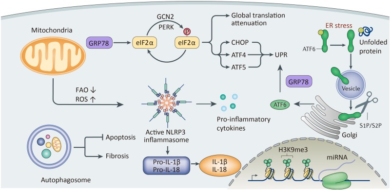
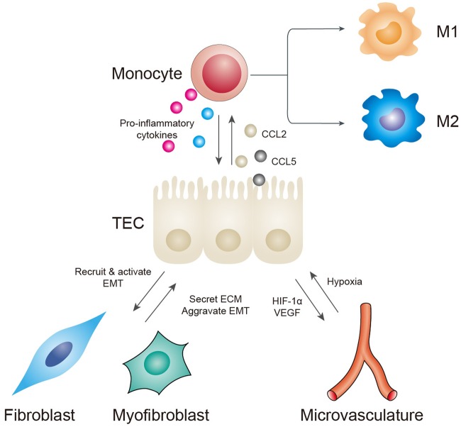
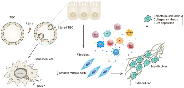
Renal fibrosis, especially tubulointerstitial fibrosis, is the inevitable outcome of all progressive chronic kidney diseases (CKDs) and exerts a great health burden worldwide. For a long time, interests in renal fibrosis have been concentrated on fibroblasts and myofibroblasts. However, in recent years, growing numbers of studies have focused on the role of tubular epithelial cells (TECs). TECs, rather than a victim or bystander, are probably a neglected mediator in renal fibrosis, responding to a variety of injuries. The maladaptive repair mechanisms of TECs may be the key point in this process. In this review, we will focus on the role of TECs in tubulointerstitial fibrosis. We will follow the fate of a tubular cell and depict the intracellular changes after injury. We will then discuss how the repair mechanism of tubular cells becomes maladaptive, and we will finally discuss the intercellular crosstalk in the interstitium that ultimately proceeds tubulointerstitial fibrosis.

摘要

肾纤维化,尤其是肾小管间质纤维化,是所有进行性慢性肾脏病(CKD)的必然结局,在全球范围内造成了巨大的健康负担。长期以来,人们对肾纤维化的兴趣主要集中在成纤维细胞和肌成纤维细胞上。然而,近年来,越来越多的研究关注肾小管上皮细胞(TECs)的作用。TECs 不是受害者或旁观者,而是肾纤维化中被忽视的重要介质,对各种损伤作出反应。TECs 的适应性修复机制可能是这一过程的关键点。在这篇综述中,我们将重点关注 TECs 在肾小管间质纤维化中的作用。我们将跟随一个肾小管细胞的命运,描述损伤后的细胞内变化。然后,我们将讨论肾小管细胞的修复机制如何变得失调,最后,我们将讨论间质中的细胞间串扰,最终导致肾小管间质纤维化。

https://cdn.ncbi.nlm.nih.gov/pmc/blobs/7748/6233178/5e7b9e88ece0/41419_2018_1157_Fig3_HTML.jpg
https://cdn.ncbi.nlm.nih.gov/pmc/blobs/7748/6233178/11f96cea7fb0/41419_2018_1157_Fig1_HTML.jpg
https://cdn.ncbi.nlm.nih.gov/pmc/blobs/7748/6233178/f4c13a56fa8f/41419_2018_1157_Fig2_HTML.jpg
https://cdn.ncbi.nlm.nih.gov/pmc/blobs/7748/6233178/5e7b9e88ece0/41419_2018_1157_Fig3_HTML.jpg
https://cdn.ncbi.nlm.nih.gov/pmc/blobs/7748/6233178/11f96cea7fb0/41419_2018_1157_Fig1_HTML.jpg
https://cdn.ncbi.nlm.nih.gov/pmc/blobs/7748/6233178/f4c13a56fa8f/41419_2018_1157_Fig2_HTML.jpg
https://cdn.ncbi.nlm.nih.gov/pmc/blobs/7748/6233178/5e7b9e88ece0/41419_2018_1157_Fig3_HTML.jpg

相似文献

1
Renal tubular epithelial cells: the neglected mediator of tubulointerstitial fibrosis after injury.肾管状上皮细胞:损伤后小管间质性纤维化的被忽视的中介。
Cell Death Dis. 2018 Nov 13;9(11):1126. doi: 10.1038/s41419-018-1157-x.
2
Distinct T cell/renal tubular epithelial cell interactions define differential chemokine production: implications for tubulointerstitial injury in chronic glomerulonephritides.独特的T细胞/肾小管上皮细胞相互作用决定了趋化因子产生的差异:对慢性肾小球肾炎肾小管间质损伤的影响
J Immunol. 2000 Mar 15;164(6):3323-9. doi: 10.4049/jimmunol.164.6.3323.
3
Inflammasome-independent NLRP3 augments TGF-β signaling in kidney epithelium.NLRP3 炎症小体非依赖性增强肾脏上皮细胞中的 TGF-β 信号转导。
J Immunol. 2013 Feb 1;190(3):1239-49. doi: 10.4049/jimmunol.1201959. Epub 2012 Dec 21.
4
The C5a/C5aR1 axis promotes progression of renal tubulointerstitial fibrosis in a mouse model of renal ischemia/reperfusion injury.C5a/C5aR1 轴促进了肾缺血/再灌注损伤小鼠模型肾小管间质纤维化的进展。
Kidney Int. 2019 Jul;96(1):117-128. doi: 10.1016/j.kint.2019.01.039. Epub 2019 Mar 4.
5
HIF-1α inducing exosomal microRNA-23a expression mediates the cross-talk between tubular epithelial cells and macrophages in tubulointerstitial inflammation.缺氧诱导因子-1α诱导的外泌体 microRNA-23a 表达介导肾小管上皮细胞与巨噬细胞之间在肾小管间质性炎症中的串扰。
Kidney Int. 2019 Feb;95(2):388-404. doi: 10.1016/j.kint.2018.09.013. Epub 2018 Dec 11.
6
[Identification of Biomarkers for Tubular Injury and Interstitial Fibrosis in Chronic Kidney Disease].[慢性肾脏病肾小管损伤和间质纤维化生物标志物的鉴定]
Yakugaku Zasshi. 2017;137(11):1355-1360. doi: 10.1248/yakushi.17-00150.
7
Actin filaments in human renal tubulo-interstitial fibrosis: significance for the concept of epithelial-myofibroblast transformation.人肾小管间质纤维化中的肌动蛋白丝:对上皮-肌成纤维细胞转化概念的意义
J Submicrosc Cytol Pathol. 2003 Jul;35(3):221-33.
8
Inflammatory cell infiltration and resolution of kidney inflammation is orchestrated by the cold-shock protein Y-box binding protein-1.炎症细胞浸润和肾脏炎症的消退是由冷休克蛋白 Y 盒结合蛋白-1 协调的。
Kidney Int. 2017 Nov;92(5):1157-1177. doi: 10.1016/j.kint.2017.03.035. Epub 2017 Jun 11.
9
Compound K inhibits priming and mitochondria-associated activating signals of NLRP3 inflammasome in renal tubulointerstitial lesions.化合物 K 抑制 NLRP3 炎性小体在肾小管间质性病变中的引发和线粒体相关激活信号。
Nephrol Dial Transplant. 2020 Jan 1;35(1):74-85. doi: 10.1093/ndt/gfz073.
10
Protease-activated receptor 2 does not contribute to renal inflammation or fibrosis in the obstructed kidney.蛋白酶激活受体 2 不参与梗阻肾脏的炎症或纤维化反应。
Nephrology (Carlton). 2019 Sep;24(9):983-991. doi: 10.1111/nep.13635. Epub 2019 Jul 31.

引用本文的文献

1
Inhibition of the YAP-E2F2-FGF2 axis in renal tubular cells ameliorates renal fibrosis in chronic kidney disease.抑制肾小管细胞中的YAP-E2F2-FGF2轴可改善慢性肾脏病中的肾纤维化。
Sci China Life Sci. 2025 Sep 10. doi: 10.1007/s11427-024-2880-1.
2
Loss of PCAF in proximal tubular cells exacerbates renal fibrosis by promoting partial epithelial-to-mesenchymal transition.近端肾小管细胞中PCAF的缺失通过促进部分上皮-间充质转化加剧肾纤维化。
Exp Mol Med. 2025 Sep 1. doi: 10.1038/s12276-025-01533-x.
3
Comparative Analysis of Curcumin and Metformin Effects on the Kidney in a Model of Unilateral Nephrectomy and Ischemia-Reperfusion Injury.

本文引用的文献

1
Membrane rafts-redox signalling pathway contributes to renal fibrosis via modulation of the renal tubular epithelial-mesenchymal transition.膜筏-氧化还原信号通路通过调节肾小管上皮-间充质转化参与肾纤维化。
J Physiol. 2018 Aug;596(16):3603-3616. doi: 10.1113/JP275952. Epub 2018 Jul 23.
2
Acute Kidney Injury as a Condition of Renal Senescence.急性肾损伤作为肾脏衰老的一种情况。
Cell Transplant. 2018 May;27(5):739-753. doi: 10.1177/0963689717743512. Epub 2018 Apr 27.
3
CD4 and CD8 T Cells Exert Regulatory Properties During Experimental Acute Aristolochic Acid Nephropathy.
姜黄素与二甲双胍对单侧肾切除及缺血再灌注损伤模型肾脏影响的比较分析
Rep Biochem Mol Biol. 2025 Jan;13(4):579-589. doi: 10.61186/rbmb.13.4.579.
4
Injured tubule derived CCN1 exacerbates renal congestion-mediated acute kidney injury and fibrosis.损伤肾小管来源的CCN1会加重肾淤血介导的急性肾损伤和纤维化。
Sci Rep. 2025 Jul 1;15(1):20840. doi: 10.1038/s41598-025-05723-5.
5
and Flower Extract Complex Alleviates Kidney Inflammation and Fibrosis by Modulating Oxidative Stress.花提取物复合物通过调节氧化应激减轻肾脏炎症和纤维化。
Antioxidants (Basel). 2025 Mar 28;14(4):409. doi: 10.3390/antiox14040409.
6
Association between uric acid to high-density lipoprotein cholesterol ratio and chronic kidney disease in Chinese patients with type 2 diabetes mellitus: a cross-sectional study.中国 2 型糖尿病患者尿酸与高密度脂蛋白胆固醇比值和慢性肾脏病的相关性:一项横断面研究
Front Nutr. 2025 Apr 14;12:1582495. doi: 10.3389/fnut.2025.1582495. eCollection 2025.
7
Renal tubular epithelial cell-derived Exosomal miR-330-3p plays a key role in fibroblast activation and renal fibrosis by regulating CREBBP.肾小管上皮细胞衍生的外泌体miR-330-3p通过调节CREBBP在成纤维细胞活化和肾纤维化中起关键作用。
Stem Cell Res Ther. 2025 Apr 23;16(1):203. doi: 10.1186/s13287-025-04338-x.
8
Injured tubular epithelia-derived CCN1 promotes the mobilization of fibroblasts toward injury sites after kidney injury.受损的肾小管上皮细胞衍生的CCN1促进肾损伤后成纤维细胞向损伤部位的动员。
iScience. 2025 Mar 7;28(4):112176. doi: 10.1016/j.isci.2025.112176. eCollection 2025 Apr 18.
9
Acetylation-regulated DUSP1 deficiency contributes to renal fibrosis progression.乙酰化调控的双特异性磷酸酶1缺乏促进肾纤维化进展。
Theranostics. 2025 Mar 3;15(9):3781-3796. doi: 10.7150/thno.108992. eCollection 2025.
10
Emo@KP MBs Modulates the TGF-β1/Smad Signaling Pathway by in situ Micro-Nano Conversion to Reduce Renal Inflammation and Fibrosis Caused by Unilateral Ureteral Obstruction.Emo@KP微球通过原位微纳转化调节TGF-β1/Smad信号通路,减轻单侧输尿管梗阻所致的肾脏炎症和纤维化
Int J Nanomedicine. 2025 Mar 24;20:3731-3747. doi: 10.2147/IJN.S499550. eCollection 2025.
CD4 和 CD8 T 细胞在实验性急性马兜铃酸肾病中发挥调节作用。
Sci Rep. 2018 Mar 28;8(1):5334. doi: 10.1038/s41598-018-23565-2.
4
Acute Kidney Injury and Progression of Diabetic Kidney Disease.急性肾损伤与糖尿病肾病进展。
Adv Chronic Kidney Dis. 2018 Mar;25(2):166-180. doi: 10.1053/j.ackd.2017.12.005.
5
Tranilast prevents renal interstitial fibrosis by blocking mast cell infiltration in a rat model of diabetic kidney disease.曲尼司特通过阻止肥大细胞浸润预防糖尿病肾病大鼠模型的肾间质纤维化。
Mol Med Rep. 2018 May;17(5):7356-7364. doi: 10.3892/mmr.2018.8776. Epub 2018 Mar 19.
6
miR-29b regulates Ang II-induced EMT of rat renal tubular epithelial cells via targeting PI3K/AKT signaling pathway.miR-29b 通过靶向 PI3K/AKT 信号通路调节血管紧张素 II 诱导的大鼠肾小管上皮细胞 EMT。
Int J Mol Med. 2018 Jul;42(1):453-460. doi: 10.3892/ijmm.2018.3579. Epub 2018 Mar 22.
7
Specific Inhibitor of Smad3 (SIS3) Attenuates Fibrosis, Apoptosis, and Inflammation in Unilateral Ureteral Obstruction Kidneys by Inhibition of Transforming Growth Factor β (TGF-β)/Smad3 Signaling.特异性 Smad3 抑制剂(SIS3)通过抑制转化生长因子 β(TGF-β)/Smad3 信号通路减轻单侧输尿管梗阻肾脏的纤维化、细胞凋亡和炎症。
Med Sci Monit. 2018 Mar 20;24:1633-1641. doi: 10.12659/msm.909236.
8
Notch1 regulates PTEN expression to exacerbate renal tubulointerstitial fibrosis in diabetic nephropathy by inhibiting autophagy via interactions with Hes1.Notch1通过与Hes1相互作用抑制自噬来调节PTEN表达,从而加剧糖尿病肾病中的肾小管间质纤维化。
Biochem Biophys Res Commun. 2018 Mar 18;497(4):1110-1116. doi: 10.1016/j.bbrc.2018.02.187. Epub 2018 Feb 26.
9
Genome-wide analysis revealed that DZNep reduces tubulointerstitial fibrosis via down-regulation of pro-fibrotic genes.全基因组分析显示,DZNep 通过下调致纤维化基因来减少肾小管间质纤维化。
Sci Rep. 2018 Feb 28;8(1):3779. doi: 10.1038/s41598-018-22180-5.
10
Advanced Oxidation Protein Products Aggravate Tubulointerstitial Fibrosis Through Protein Kinase C-Dependent Mitochondrial Injury in Early Diabetic Nephropathy.晚期氧化蛋白产物通过蛋白激酶 C 依赖的线粒体损伤加重早期糖尿病肾病肾小管间质纤维化。
Antioxid Redox Signal. 2019 Mar 20;30(9):1162-1185. doi: 10.1089/ars.2017.7208. Epub 2018 May 2.