Landowska Aleksandra, Roberts David, Eachus Peter, Barrett Alan
Department of Psychology, School of Health Sciences, University of Salford, Salford, United Kingdom.
Military Veterans' Service, Pennine Care NHS Foundation Trust, Ashton-under-Lyne, United Kingdom.
Front Hum Neurosci. 2018 Nov 1;12:362. doi: 10.3389/fnhum.2018.00362. eCollection 2018.
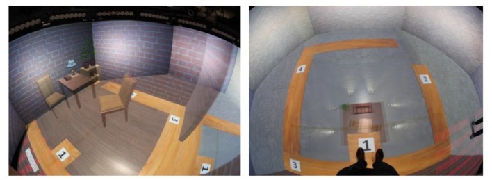
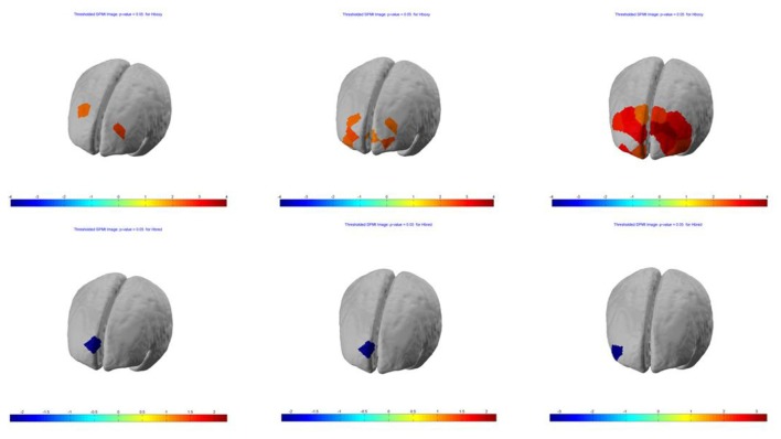
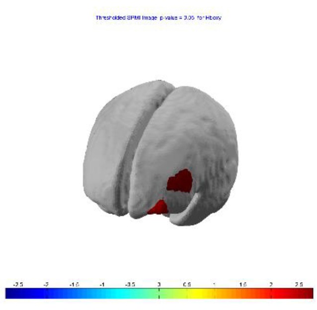
Exposure Therapy (ET) has demonstrated its efficacy in the treatment of phobias, anxiety and Post-traumatic Stress Disorder (PTSD), however, it suffers a high drop-out rate because of too low or too high patient engagement in treatment. Virtual Reality Exposure Therapy (VRET) is comparably effective regarding symptom reduction and offers an alternative tool to facilitate engagement for avoidant participants. Neuroimaging studies have demonstrated that both ET and VRET normalize brain activity within a fear circuit. However, previous studies have employed brain imaging technology which restricts people's movement and hides their body, surroundings and therapist from view. This is at odds with the way engagement is typically controlled. We used a novel combination of neural imaging and VR technology-Functional Near-Infrared Spectroscopy (fNIRS) and Immersive Projection Technology (IPT), to avoid these limitations. Although there are a few studies that have investigated the effect of VRET on a brain function after the treatment, the present study utilized technologies which promote ecological validity to measure brain changes after VRET treatment. Furthermore, there are no studies that have measured brain activity within VRET session. In this study brain activity within the prefrontal cortex (PFC) was measured during three consecutive exposure sessions. = 13 acrophobic volunteers were asked to walk on a virtual plank with a 6 m drop below. Changes in oxygenated (HbO) hemoglobin concentrations in the PFC were measured in three blocks using fNIRS. Consistent with previous functional magnetic resonance imaging (fMRI) studies, the analysis showed decreased activity in the DLPFC and MPFC during first exposure. The activity increased toward normal across three sessions. The study demonstrates potential efficacy of a method for measuring within-session neural response to virtual stimuli that could be replicated within clinics and research institutes, with equipment better suited to an ET session and at fraction of the cost, when compared to fMRI. This has application in widening access to, and increasing ecological validity of, immersive neuroimaging across understanding, diagnosis, assessment and treatment of, a range of mental disorders such as phobia, anxiety and PTSD or addictions.
暴露疗法(ET)已在恐惧症、焦虑症和创伤后应激障碍(PTSD)的治疗中显示出其有效性,然而,由于患者对治疗的参与度过低或过高,该疗法的退出率很高。虚拟现实暴露疗法(VRET)在减轻症状方面具有同等效果,并为促进回避型参与者的参与提供了一种替代工具。神经影像学研究表明,ET和VRET都能使恐惧回路中的大脑活动正常化。然而,以前的研究采用的脑成像技术限制了人们的行动,并将他们的身体、周围环境和治疗师隐藏起来,使其无法被看到。这与通常控制参与度的方式不一致。我们使用了神经成像和VR技术的新组合——功能近红外光谱(fNIRS)和沉浸式投影技术(IPT),以避免这些限制。尽管有一些研究调查了VRET治疗后对脑功能的影响,但本研究采用了提高生态效度的技术来测量VRET治疗后的大脑变化。此外,还没有研究测量过VRET治疗过程中的大脑活动。在本研究中,在连续三次暴露治疗过程中测量了前额叶皮层(PFC)的大脑活动。13名恐高症志愿者被要求在下方有6米落差的虚拟木板上行走。使用fNIRS在三个时间段测量PFC中氧合血红蛋白(HbO)浓度的变化。与之前的功能磁共振成像(fMRI)研究一致,分析显示在第一次暴露期间,背外侧前额叶皮层(DLPFC)和内侧前额叶皮层(MPFC)的活动减少。在三个治疗阶段中,活动逐渐恢复到正常水平。该研究证明了一种测量对虚拟刺激的治疗过程中神经反应方法的潜在有效性,与fMRI相比,该方法可以在诊所和研究机构中复制,使用更适合暴露疗法治疗过程的设备,且成本仅为其一小部分。这在扩大对一系列精神障碍(如恐惧症、焦虑症和创伤后应激障碍或成瘾)的理解、诊断、评估和治疗中沉浸式神经成像的可及性和提高其生态效度方面具有应用价值。