Department of Paediatrics, The Hospital for Sick Children, 555 University Avenue, Toronto, ON M5G 1X8, Canada; Faculty of Education, The University of Western Ontario, 1137 Western Rd, London, ON, N6G 1G7, Canada.
Department of Paediatrics, The Hospital for Sick Children, 555 University Avenue, Toronto, ON M5G 1X8, Canada.
Neuroimage Clin. 2019;21:101596. doi: 10.1016/j.nicl.2018.11.006. Epub 2018 Nov 13.
To determine whether the spatial extent and location of early-identified punctate white matter injury (WMI) is associated with regionally-specific disruptions in thalamocortical-connectivity in very-preterm born neonates.
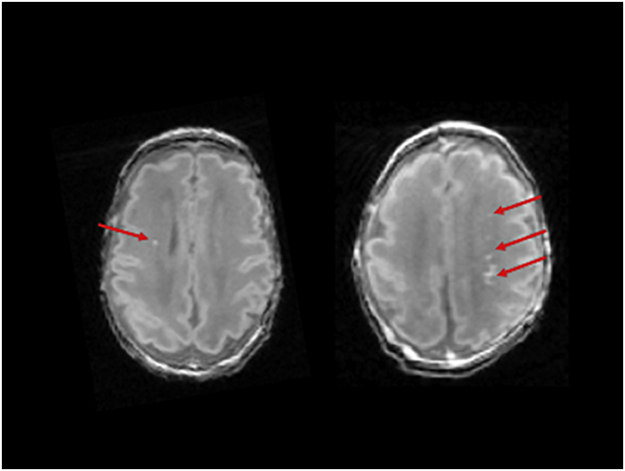
37 very-preterm born neonates (median gestational age: 28.1 weeks; interquartile range [IQR]: 27-30) underwent early MRI (median age 32.9 weeks; IQR: 32-35), and WMI was identified in 13 (35%) neonates. Structural T1-weighted, resting-state functional Magnetic Resonance Imaging (rs-fMRI, n = 34) and Diffusion Tensor Imaging (DTI, n = 31) sequences were acquired using 3 T-MRI. A probabilistic map of WMI was developed for the 13 neonates demonstrating brain injury. A neonatal atlas was applied to the WMI maps, rs-fMRI and DTI analyses to extract volumetric, functional and microstructural data from regionally-specific brain areas. Associations of thalamocortical-network strength and alterations in fractional anisotropy (FA, a measure of white-matter microstructure) with WMI volume were assessed in general linear models, adjusting for age at scan and cerebral volumes.
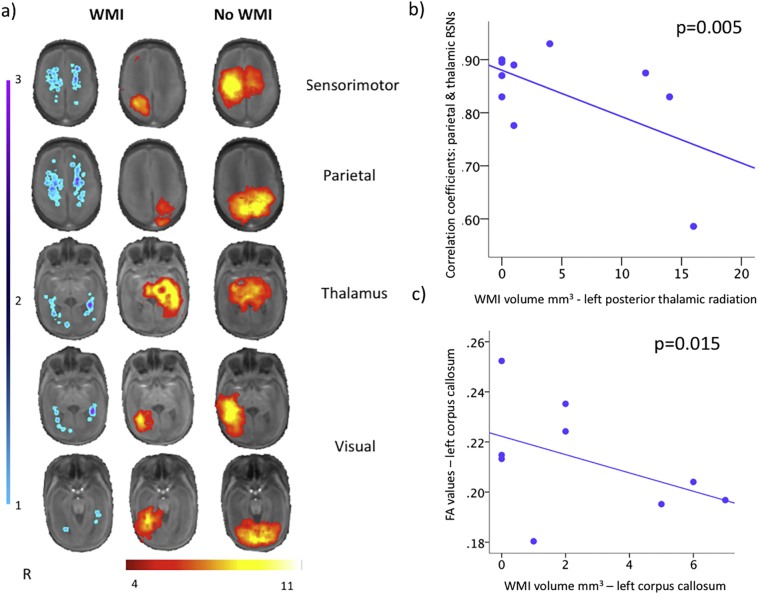
WMI volume in the superior (β = -0.007; p = .02) and posterior corona radiata (β = -0.01; p = .01), posterior thalamic radiations (β = -0.01; p = .005) and superior longitudinal fasciculus (β = -0.02; p = .001) was associated with reduced connectivity strength between thalamus and parietal resting-state networks. WMI volume in the left (β = -0.02; p = .02) and right superior corona radiata (β = -0.03; p = .008), left posterior corona radiata (β = -0.03; p = .01), corpus callosum (β = -0.11; p < .0001) and right superior longitudinal fasciculus (β = -0.02; p = .02) was associated with functional connectivity strength between thalamic and sensorimotor networks. Increased WMI volume was also associated with decreased FA values in the corpus callosum (β = -0.004, p = .015).
Regionally-specific alterations in early functional and structural network complexity resulting from WMI may underlie impaired outcomes.
确定早期识别的点状脑白质损伤(WMI)的空间范围和位置是否与极早产儿丘脑皮质连接的区域性特异性中断有关。
37 名极早产儿(中位胎龄:28.1 周;四分位距 [IQR]:27-30)接受了早期 MRI(中位年龄 32.9 周;IQR:32-35),13 名(35%)早产儿发现有 WMI。使用 3T-MRI 采集结构 T1 加权、静息态功能磁共振成像(rs-fMRI,n=34)和弥散张量成像(DTI,n=31)序列。为显示脑损伤的 13 名新生儿绘制了 WMI 概率图。将新生儿图谱应用于 WMI 图谱、rs-fMRI 和 DTI 分析,从区域特异性脑区提取容积、功能和微观结构数据。在一般线性模型中评估了丘脑皮质网络强度与各向异性分数(FA,白质微观结构的度量)与 WMI 体积之间的关联,调整了扫描时的年龄和脑容量。
在优势(β= -0.007;p=.02)和后冠状辐射(β= -0.01;p=.01)、后丘脑辐射(β= -0.01;p=.005)和上纵束(β= -0.02;p=.001)的 WMI 体积与丘脑与顶叶静息状态网络之间的连接强度降低有关。左侧(β= -0.02;p=.02)和右侧优势冠状辐射(β= -0.03;p=.008)、左侧后冠状辐射(β= -0.03;p=.01)、胼胝体(β= -0.11;p<.0001)和右侧上纵束(β= -0.02;p=.02)的 WMI 体积与丘脑与感觉运动网络之间的功能连接强度有关。WMI 体积增加也与胼胝体 FA 值降低有关(β= -0.004,p=.015)。
WMI 导致的早期功能和结构网络复杂性的区域性特异性改变可能是导致预后不良的原因。