Capobres Thomas, Sabharwal Gauravi, Griffith Brent
Department of Radiology, Henry Ford Health System, Detroit, MI, USA.
BJR Case Rep. 2016 Jul 28;2(3):20150420. doi: 10.1259/bjrcr.20150420. eCollection 2016.
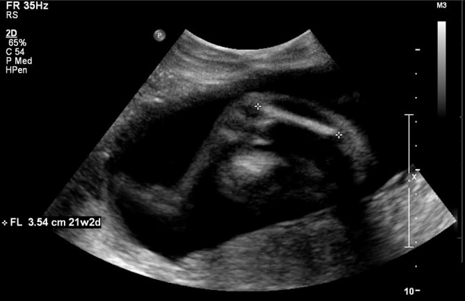
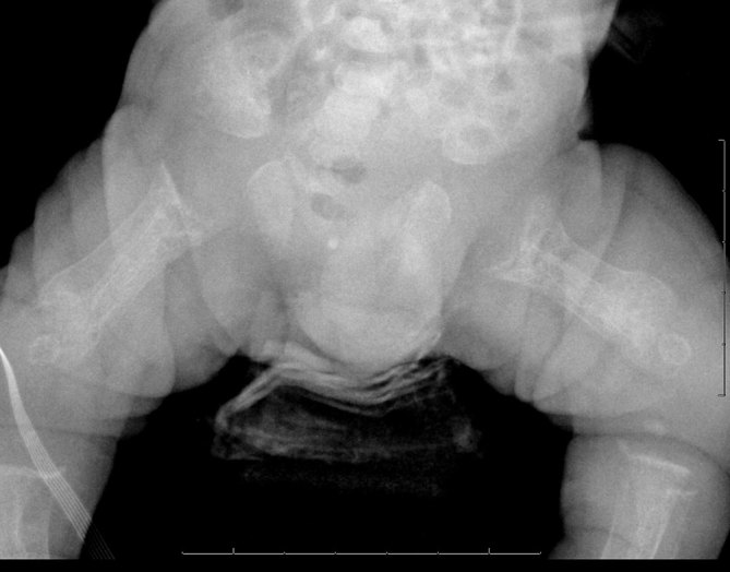
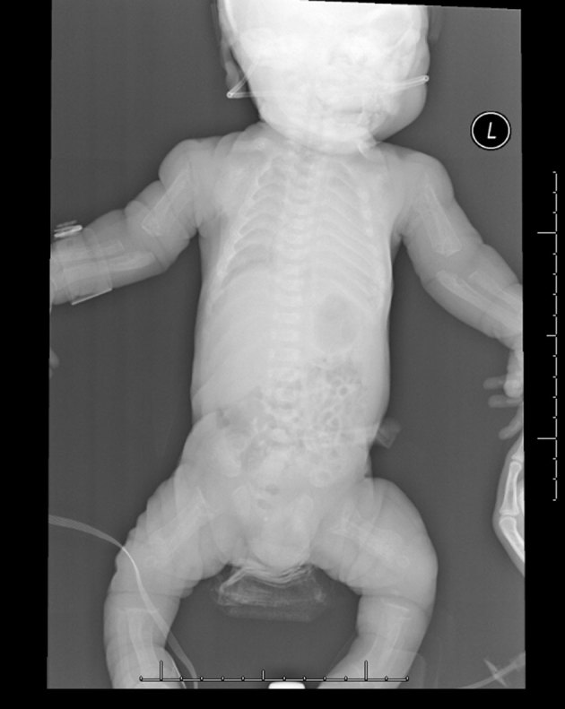
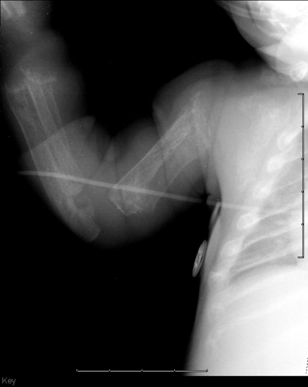
A 28-year-old G3 P1 SAB1 female with no prior health concerns was found to have a foetus with short femurs on prenatal ultrasound following an abnormal maternal serum screen result. Fluid obtained by amniocentesis revealed an elevated α-fetoprotein level with absence of an acetylcholinesterase band and normal male karyotype (46,XY). Follow-up ultrasound 3 weeks later again demonstrated short femur lengths, but no other abnormalities. At birth, the child was noted to have multiple dysmorphic features, including short humeri and femurs, coarse facial features, retrognathia and yellowish hypertrophic gums in addition to hyperbilirubinaemia and thrombocytopenia. Radiological studies demonstrated bony demineralization with profound diaphyseal cloaking in the long bones. Genetic testing diagnosed I-cell disease.
一名28岁、孕3产1、有1次自然流产史的女性,既往无健康问题,在母体血清筛查结果异常后,产前超声检查发现胎儿股骨短小。羊水穿刺获得的液体显示甲胎蛋白水平升高,无乙酰胆碱酯酶带,核型为正常男性(46,XY)。3周后的随访超声检查再次显示股骨长度短,但无其他异常。出生时,该患儿有多种畸形特征,包括肱骨和股骨短小、面部特征粗糙、下颌后缩、牙龈黄染肥厚,此外还有高胆红素血症和血小板减少症。放射学研究显示长骨骨质脱矿,骨干有明显的骨膜增厚。基因检测诊断为I型细胞病。