Department of Orthopedics, Affiliated Traditional Chinese Medicine Hospital of Southwest Medical University, Luzhou, Sichuan, China (mainland).
Academician Workstation in Luzhou, Luzhou, Sichuan, China (mainland).
Med Sci Monit. 2018 Nov 22;24:8417-8421. doi: 10.12659/MSM.911534.
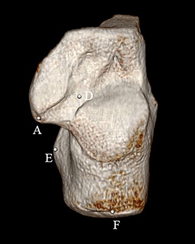
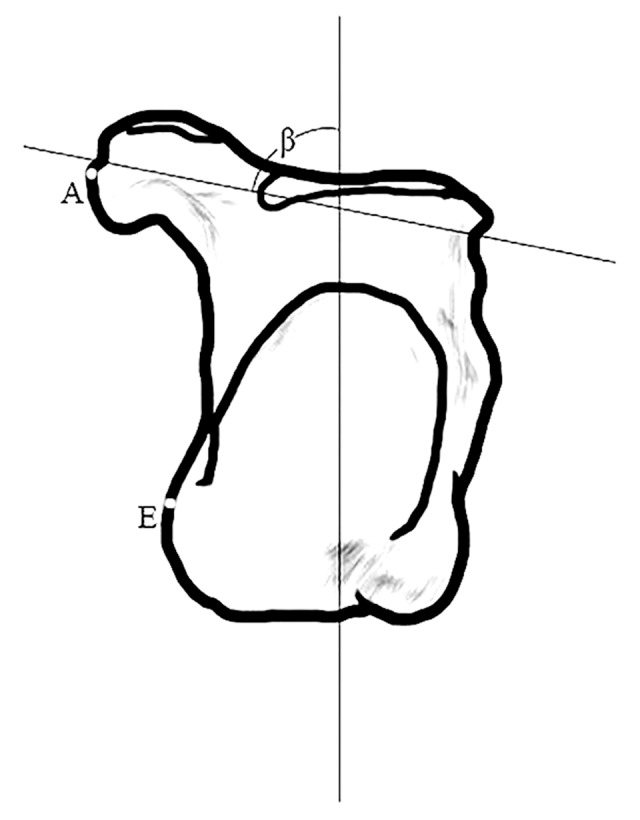
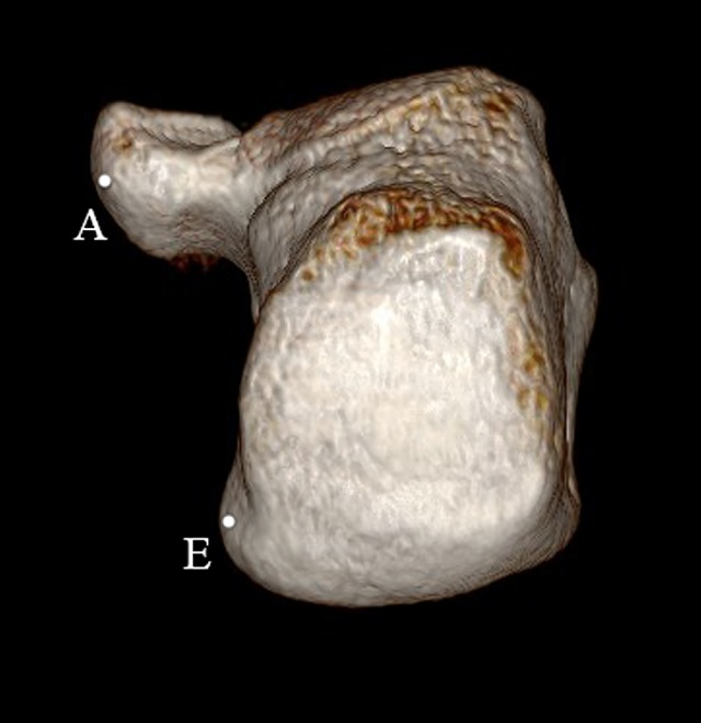
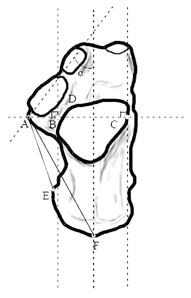
BACKGROUND With the complexity of calcaneal fracture (CF) increasing, its treatment has changed to include inserting the screw used to secure the facies articular posterior into the sustentaculum tail (ST). Some research progress has been made in this area, but there has been little in-depth research on the anatomical morphology of the sustentaculum tail, which is necessary for clinical surgery, and more information about Chinese anatomic characteristics and improved surgical techniques for CF are needed. MATERIAL AND METHODS This anatomical study, based on a three-dimensional (3D) computed tomographic (CT) reconstruction technique, included 287 dry calcaneus, consisting of 144 left and 143 right calcaneus. The images were reconstructed in 3D after CT scanning. Seven subjects were enrolled (L and R): (1) The vertical distance from inside the sustentaculum tail (IST) to inside the facies articularis talaris posterior; (2) The vertical distance from IST to the outside facies articularis talaris posterior; (3) The thickness of sulcus calcaneal nadir; (4) The distance from IST to processus medislis tuberis calcaneus; (5) The distance from IST to calcaneal posterosuperior tuber; (6) The angle of the prolate axial intersection between ST and calcaneus on the normal superior as ˂α; and (7) The angle of the prolate axial intersection between ST and calcaneus on the normal posterior as ˂β. All measurement results were analyzed by SPSS 22.0. RESULTS Based on morphological classification, the average length of AB, AC, AE, and AF on left ST were 16.956±1.391 mm, 37.803±2.525 mm, 43.244±3.617 mm, and 51.113±4.455 mm, respectively. Among the others, ˂β was 81.227±6.317 mm on the left and 74.581±9.008 mm on the right (P<0.05). CONCLUSIONS These results suggest better ways to treat the special characteristics and to reduce the risk of CF surgery.
随着跟骨骨折(CF)复杂性的增加,其治疗方法已改为将用于固定关节后关节面的螺钉插入跟骨距骨尾部(ST)。在这一领域已经取得了一些研究进展,但对跟骨距骨尾部的解剖形态研究甚少,而这对于临床手术是必要的,并且需要更多关于中国解剖特征的信息和改进的 CF 手术技术。
本解剖研究基于三维(3D)计算机断层扫描(CT)重建技术,共纳入 287 例干跟骨,包括 144 例左侧跟骨和 143 例右侧跟骨。CT 扫描后对图像进行 3D 重建。共纳入 7 名受试者(L 和 R):(1)距骨尾部内侧面(IST)到距骨后关节面的垂直距离;(2)IST 到距骨后关节面外侧的垂直距离;(3)跟骨底切迹的厚度;(4)IST 到跟骨内侧结节的距离;(5)IST 到跟骨后上结节的距离;(6)正常上位时 ST 与跟骨之间的长轴相交的角度<α;(7)正常后位时 ST 与跟骨之间的长轴相交的角度<β。所有测量结果均采用 SPSS 22.0 进行分析。
基于形态学分类,左侧 ST 的 AB、AC、AE 和 AF 的平均长度分别为 16.956±1.391mm、37.803±2.525mm、43.244±3.617mm 和 51.113±4.455mm。其中,左侧<β为 81.227±6.317mm,右侧为 74.581±9.008mm(P<0.05)。
这些结果为治疗 CF 提供了更好的方法,可降低手术风险。