Immunology and Reproduction Biology Laboratory & State Key Laboratory of Analytical Chemistry for Life Science, Medical School, Nanjing University, Hankou Road 22, Nanjing, 210093, China.
Jiangsu Key Laboratory of Molecular Medicine, Nanjing University, Nanjing, 210093, China.
Cell Commun Signal. 2018 Nov 23;16(1):89. doi: 10.1186/s12964-018-0300-8.
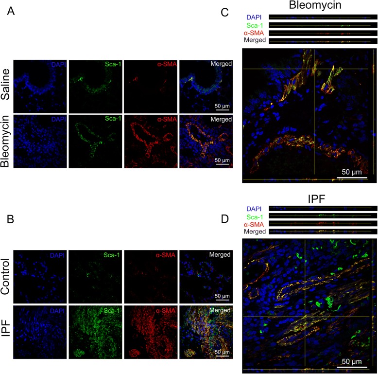
Idiopathic pulmonary fibrosis (IPF) is a devastating disease characterized by the histopathological pattern of usual interstitial pneumonia and is associated with a high mortality rate. Recently, lung resident mesenchymal stem cells (LR-MSCs) have been identified as an important contributor to myofibroblast activation in pulmonary fibrosis. Macrophages are also believed to play a critical role in pulmonary fibrosis. However, the underlying connections between LR-MSCs and macrophages in the pathogenesis of pulmonary fibrosis are still elusive.
In this study, we investigated the interaction between LR-MSCs and macrophages using a bleomycin-induced mouse pulmonary fibrosis model and a coculture system.
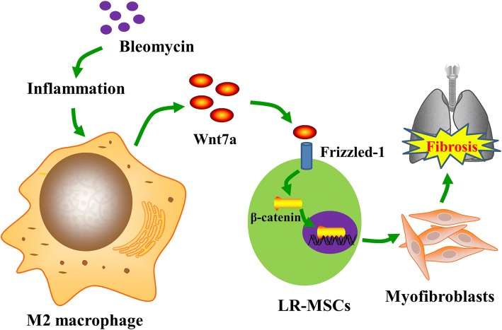
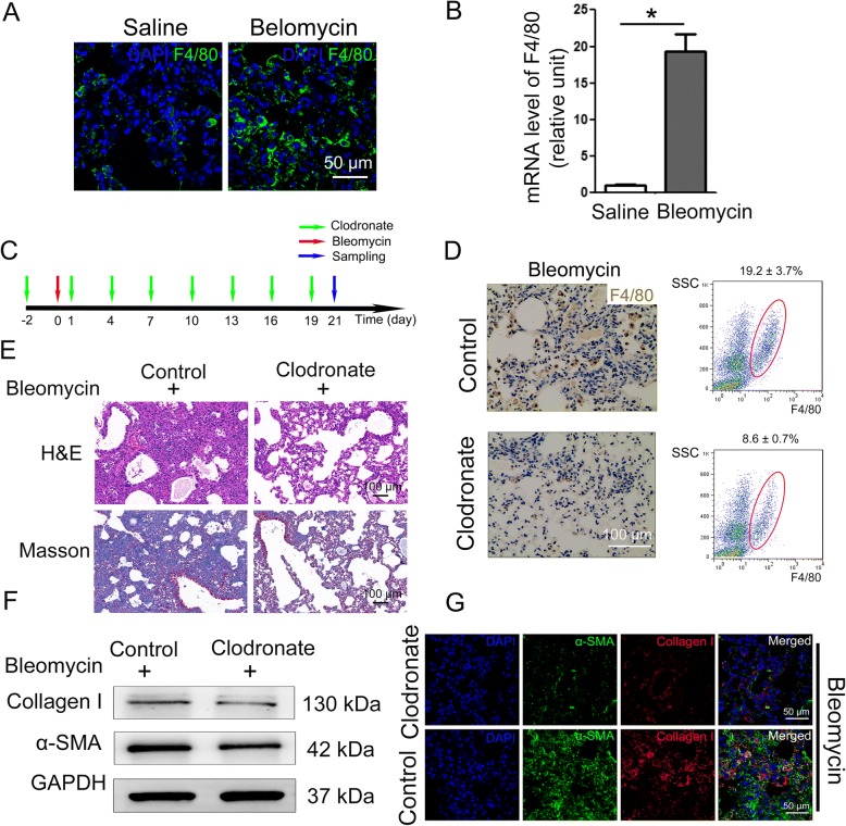
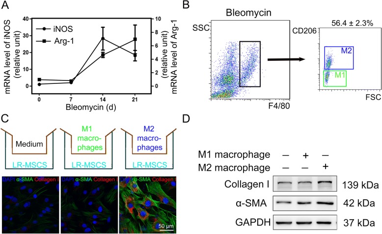
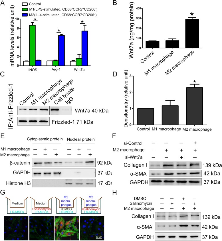
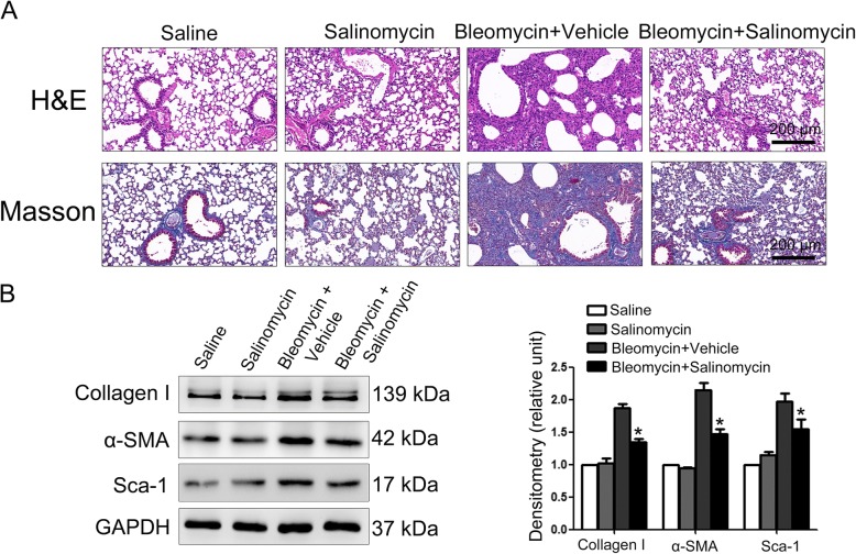
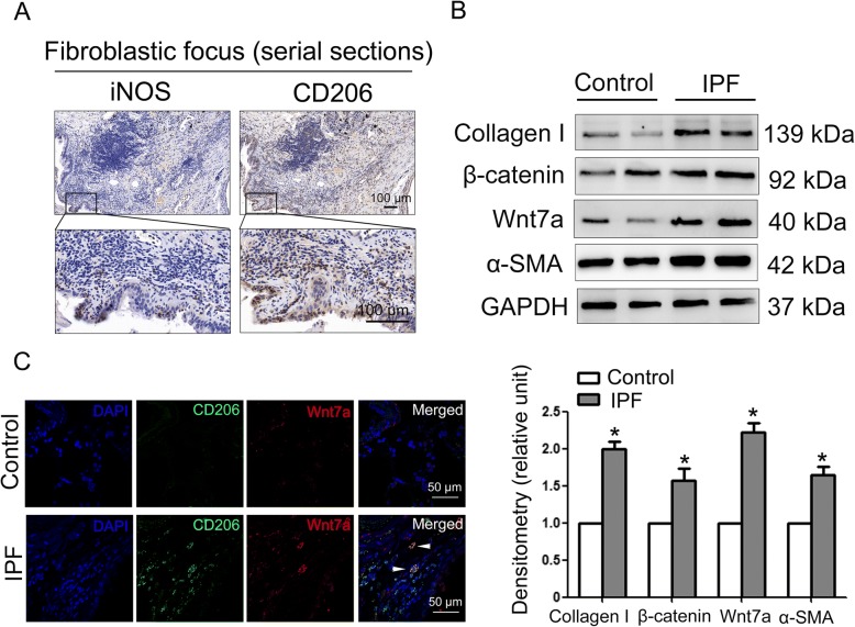
Here, we show that blocking pulmonary macrophage infiltration attenuated bleomycin-induced pulmonary fibrosis. In addition, as determined by flow cytometry, we discovered that the recruited macrophages in fibrotic lungs of bleomycin-treated mice were mainly M2 macrophages. In particular, we found that M2, rather than M1 macrophages, promoted myofibroblast differentiation of LR-MSCs. Moreover, we demonstrated that suppression of the Wnt/β-catenin signaling pathway could attenuate myofibroblast differentiation of LR-MSCs induced by M2 macrophages and bleomycin-induced pulmonary fibrosis. Tissue samples from IPF patients confirmed the infiltration of M2 macrophages and activation of Wnt/β-catenin signaling pathway.
In summary, this study furthered our understanding of the pulmonary fibrosis pathogenesis and highlighted M2 macrophages as a critical target for treating pulmonary fibrosis.
特发性肺纤维化(IPF)是一种破坏性疾病,其组织病理学表现为寻常型间质性肺炎,死亡率较高。最近,肺固有间充质干细胞(LR-MSCs)被认为是肺纤维化中肌成纤维细胞激活的重要贡献者。巨噬细胞也被认为在肺纤维化中发挥关键作用。然而,LR-MSCs 和巨噬细胞在肺纤维化发病机制中的潜在联系仍不清楚。
在这项研究中,我们使用博来霉素诱导的小鼠肺纤维化模型和共培养系统研究了 LR-MSCs 和巨噬细胞之间的相互作用。
我们发现,阻断肺巨噬细胞浸润可减轻博来霉素诱导的肺纤维化。此外,通过流式细胞术检测,我们发现博来霉素处理的小鼠纤维化肺中的募集巨噬细胞主要为 M2 巨噬细胞。特别是,我们发现 M2 巨噬细胞,而不是 M1 巨噬细胞,促进了 LR-MSCs 的成肌纤维细胞分化。此外,我们证明抑制 Wnt/β-catenin 信号通路可以减弱 M2 巨噬细胞和博来霉素诱导的肺纤维化诱导的 LR-MSCs 成肌纤维细胞分化。来自 IPF 患者的组织样本证实了 M2 巨噬细胞的浸润和 Wnt/β-catenin 信号通路的激活。
综上所述,这项研究进一步了解了肺纤维化的发病机制,并强调了 M2 巨噬细胞作为治疗肺纤维化的一个关键靶点。