• 文献检索
  • 文档翻译
  • 深度研究
  • 学术资讯
  • Suppr Zotero 插件Zotero 插件
  • 邀请有礼
  • 套餐&价格
  • 历史记录
应用&插件
Suppr Zotero 插件Zotero 插件浏览器插件Mac 客户端Windows 客户端微信小程序
定价
高级版会员购买积分包购买API积分包
服务
文献检索文档翻译深度研究API 文档MCP 服务
关于我们
关于 Suppr公司介绍联系我们用户协议隐私条款
关注我们

Suppr 超能文献

核心技术专利:CN118964589B侵权必究
粤ICP备2023148730 号-1Suppr @ 2026

文献检索

告别复杂PubMed语法,用中文像聊天一样搜索,搜遍4000万医学文献。AI智能推荐,让科研检索更轻松。

立即免费搜索

文件翻译

保留排版,准确专业,支持PDF/Word/PPT等文件格式,支持 12+语言互译。

免费翻译文档

深度研究

AI帮你快速写综述,25分钟生成高质量综述,智能提取关键信息,辅助科研写作。

立即免费体验

机械力通过 TGFβ 信号调节肌腱细胞外基质的组织和肌腱细胞的形态发生。

Mechanical force regulates tendon extracellular matrix organization and tenocyte morphogenesis through TGFbeta signaling.

机构信息

Department of Developmental and Cell Biology, University of California, Irvine, Irvine, United States.

Center for Regenerative Medicine, Department of Orthopaedic Surgery, Massachusetts General Hospital, Harvard Stem Cell Institute, Boston, United States.

出版信息

Elife. 2018 Nov 26;7:e38069. doi: 10.7554/eLife.38069.

DOI:10.7554/eLife.38069
PMID:30475205
原文链接:https://pmc.ncbi.nlm.nih.gov/articles/PMC6345564/
Abstract

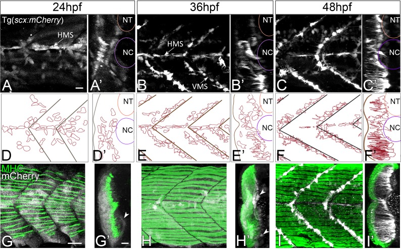
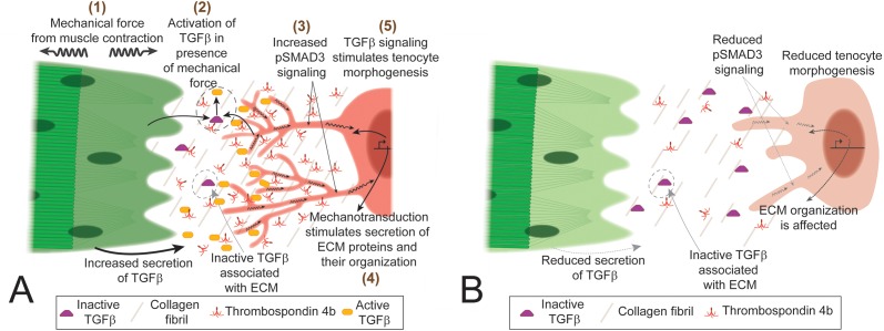
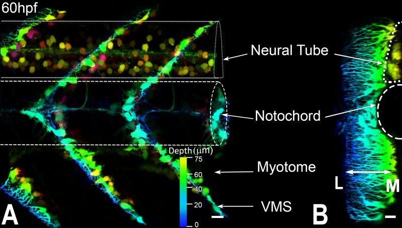
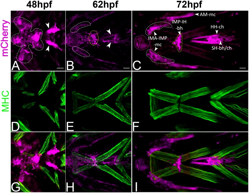
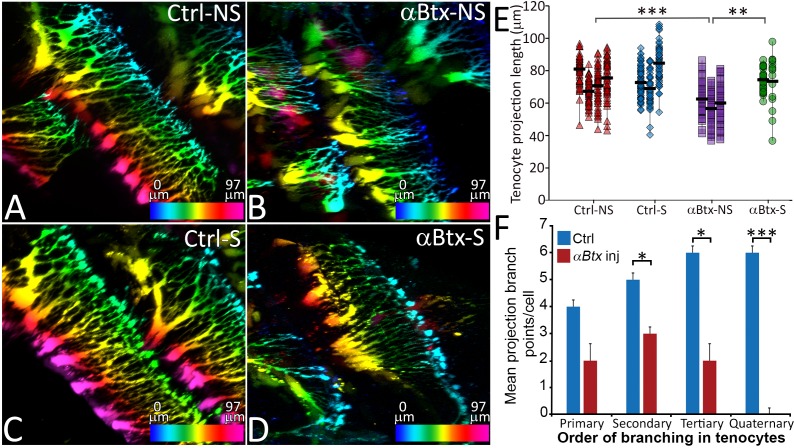
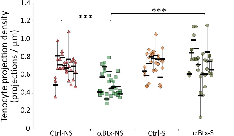
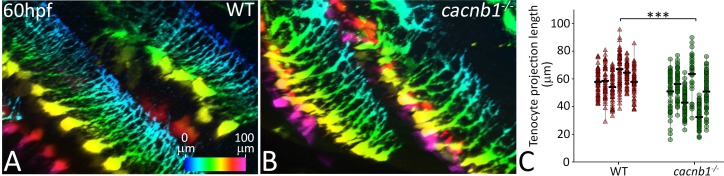
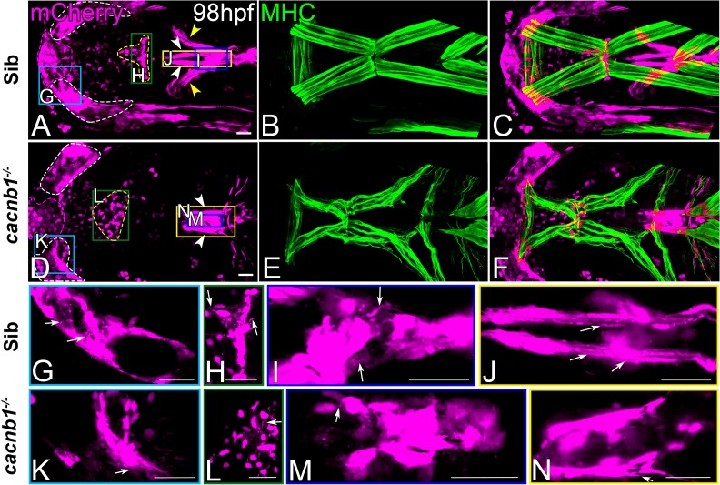
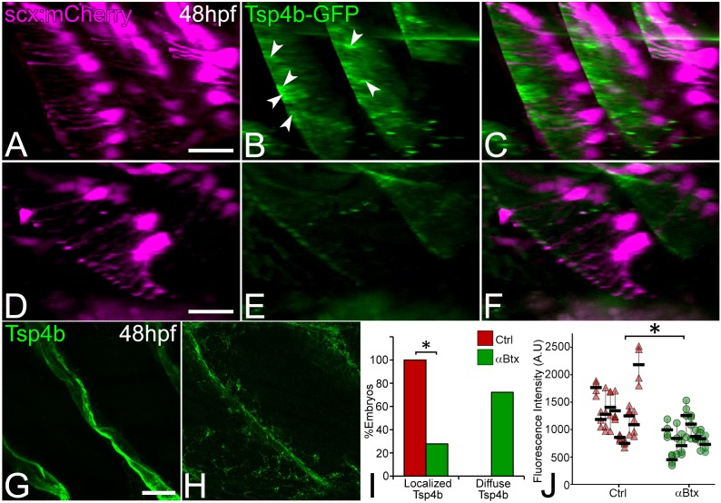
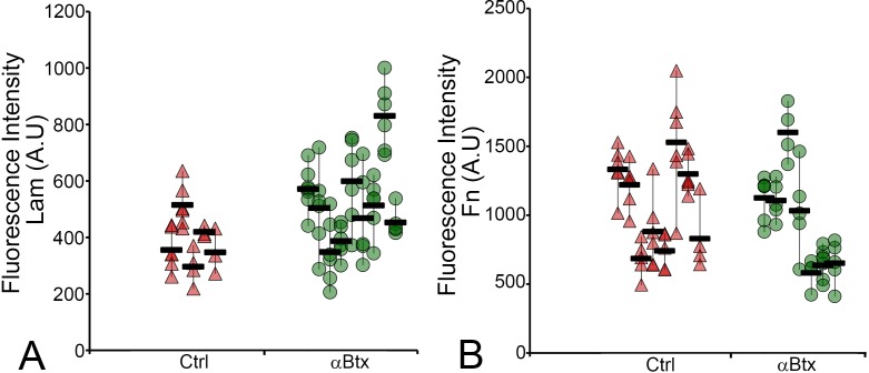
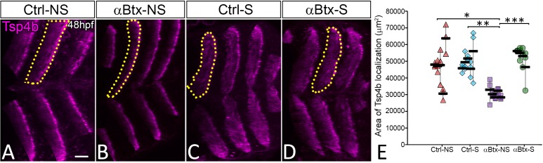
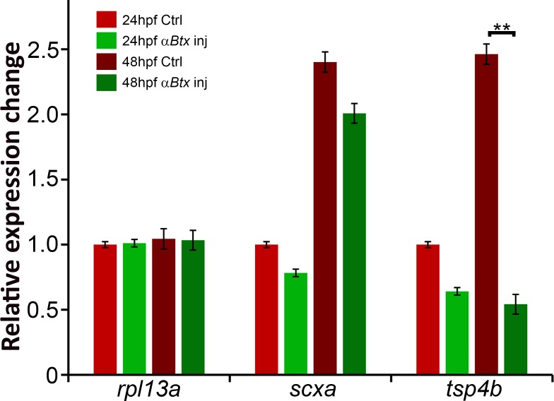
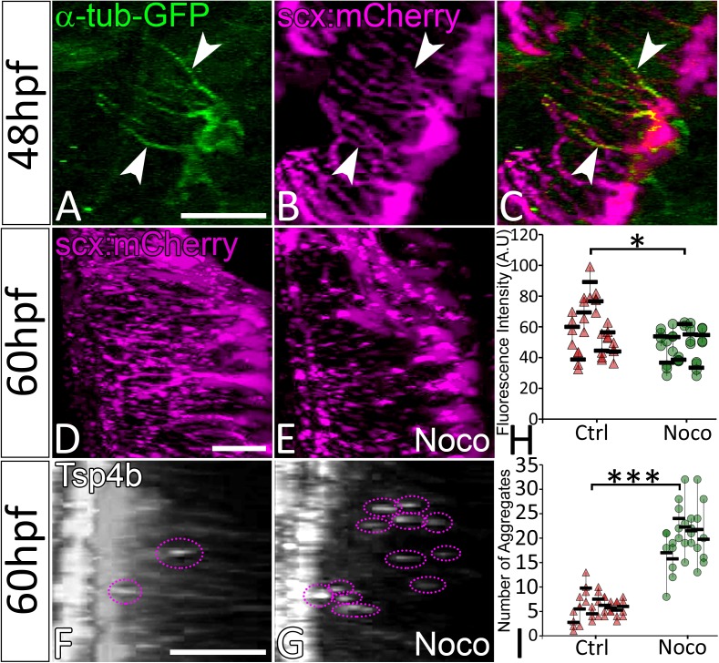
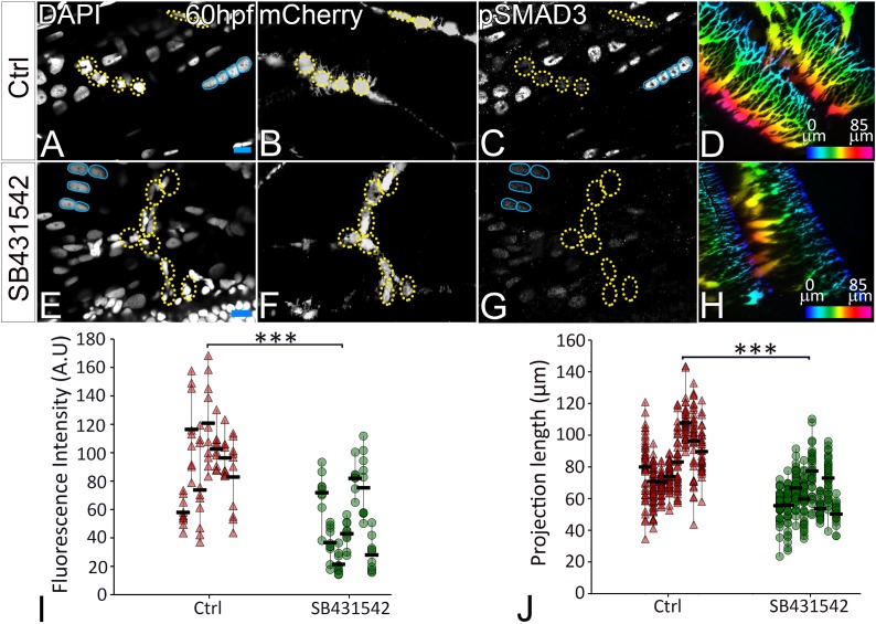
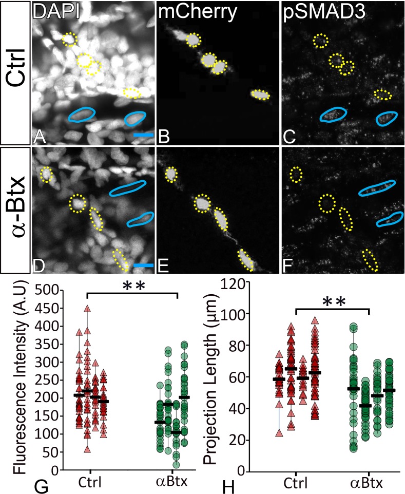
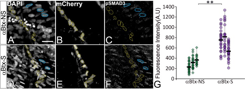
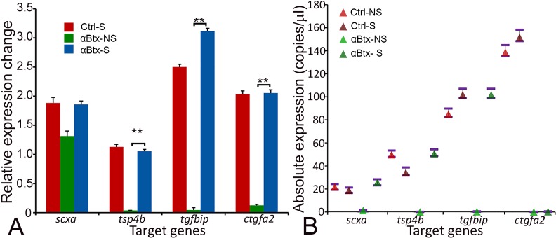
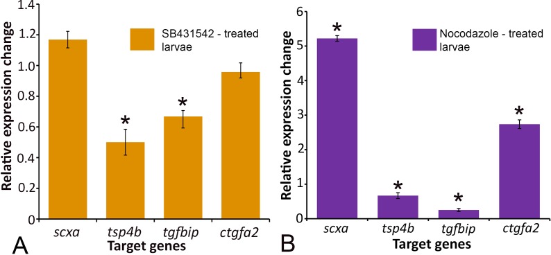
Mechanical forces between cells and extracellular matrix (ECM) influence cell shape and function. Tendons are ECM-rich tissues connecting muscles with bones that bear extreme tensional force. Analysis of transgenic zebrafish expressing mCherry driven by the tendon determinant reveals that tendon fibroblasts (tenocytes) extend arrays of microtubule-rich projections at the onset of muscle contraction. In the trunk, these form a dense curtain along the myotendinous junctions at somite boundaries, perpendicular to myofibers, suggesting a role as force sensors to control ECM production and tendon strength. Paralysis or destabilization of microtubules reduces projection length and surrounding ECM, both of which are rescued by muscle stimulation. Paralysis also reduces SMAD3 phosphorylation in tenocytes and chemical inhibition of TGFβ signaling shortens tenocyte projections. These results suggest that TGFβ, released in response to force, acts on tenocytes to alter their morphology and ECM production, revealing a feedback mechanism by which tendons adapt to tension.

摘要

细胞与细胞外基质(ECM)之间的机械力影响细胞的形状和功能。肌腱是富含细胞外基质的组织,将肌肉与承受极端张力的骨骼连接起来。对表达 mCherry 的转基因斑马鱼的分析表明,在肌肉收缩开始时,肌腱成纤维细胞(tenocytes)会延伸出一系列富含微管的突起。在躯干中,这些突起在体节边界的肌-腱连接处形成密集的幕帘,与肌纤维垂直,表明其作为力传感器的作用,以控制细胞外基质的产生和肌腱的强度。微管的麻痹或不稳定会减少突起的长度和周围的细胞外基质,肌肉刺激可以挽救这两者。麻痹还会减少 tenocytes 中的 SMAD3 磷酸化,而 TGFβ 信号的化学抑制会缩短 tenocytes 突起。这些结果表明,TGFβ 在受到力的作用时释放出来,作用于 tenocytes 以改变它们的形态和细胞外基质的产生,揭示了一种反馈机制,通过该机制,肌腱适应张力。

https://cdn.ncbi.nlm.nih.gov/pmc/blobs/b3e4/6345564/b8fd2cfff8d6/elife-38069-fig7.jpg
https://cdn.ncbi.nlm.nih.gov/pmc/blobs/b3e4/6345564/0970ddee08ae/elife-38069-fig1.jpg
https://cdn.ncbi.nlm.nih.gov/pmc/blobs/b3e4/6345564/164908f35298/elife-38069-fig1-figsupp1.jpg
https://cdn.ncbi.nlm.nih.gov/pmc/blobs/b3e4/6345564/9431b42b81f3/elife-38069-fig1-figsupp2.jpg
https://cdn.ncbi.nlm.nih.gov/pmc/blobs/b3e4/6345564/4ce88791b2b8/elife-38069-fig2.jpg
https://cdn.ncbi.nlm.nih.gov/pmc/blobs/b3e4/6345564/4d75bc6f934c/elife-38069-fig2-figsupp1.jpg
https://cdn.ncbi.nlm.nih.gov/pmc/blobs/b3e4/6345564/0bc8dfe5f8b0/elife-38069-fig2-figsupp2.jpg
https://cdn.ncbi.nlm.nih.gov/pmc/blobs/b3e4/6345564/63e156348274/elife-38069-fig2-figsupp3.jpg
https://cdn.ncbi.nlm.nih.gov/pmc/blobs/b3e4/6345564/194716557ca2/elife-38069-fig3.jpg
https://cdn.ncbi.nlm.nih.gov/pmc/blobs/b3e4/6345564/bdb089203ced/elife-38069-fig3-figsupp1.jpg
https://cdn.ncbi.nlm.nih.gov/pmc/blobs/b3e4/6345564/b14a5e5f2380/elife-38069-fig3-figsupp2.jpg
https://cdn.ncbi.nlm.nih.gov/pmc/blobs/b3e4/6345564/e96eb7ab5c3c/elife-38069-fig3-figsupp3.jpg
https://cdn.ncbi.nlm.nih.gov/pmc/blobs/b3e4/6345564/7b8e464c36e8/elife-38069-fig4.jpg
https://cdn.ncbi.nlm.nih.gov/pmc/blobs/b3e4/6345564/610b7d8eb778/elife-38069-fig5.jpg
https://cdn.ncbi.nlm.nih.gov/pmc/blobs/b3e4/6345564/3ce15354b329/elife-38069-fig6.jpg
https://cdn.ncbi.nlm.nih.gov/pmc/blobs/b3e4/6345564/f7258f23b998/elife-38069-fig6-figsupp1.jpg
https://cdn.ncbi.nlm.nih.gov/pmc/blobs/b3e4/6345564/108989c1bcf3/elife-38069-fig6-figsupp2.jpg
https://cdn.ncbi.nlm.nih.gov/pmc/blobs/b3e4/6345564/414747eac78c/elife-38069-fig6-figsupp3.jpg
https://cdn.ncbi.nlm.nih.gov/pmc/blobs/b3e4/6345564/b8fd2cfff8d6/elife-38069-fig7.jpg

相似文献

1
Mechanical force regulates tendon extracellular matrix organization and tenocyte morphogenesis through TGFbeta signaling.机械力通过 TGFβ 信号调节肌腱细胞外基质的组织和肌腱细胞的形态发生。
Elife. 2018 Nov 26;7:e38069. doi: 10.7554/eLife.38069.
2
Tendon development and musculoskeletal assembly: emerging roles for the extracellular matrix.肌腱发育与肌肉骨骼组装:细胞外基质的新作用
Development. 2015 Dec 15;142(24):4191-204. doi: 10.1242/dev.114777.
3
Thrombospondin-4 controls matrix assembly during development and repair of myotendinous junctions.血小板反应蛋白-4在肌腱-肌连接的发育和修复过程中控制基质组装。
Elife. 2014 Jun 18;3:e02372. doi: 10.7554/eLife.02372.
4
Mechanical force regulates Sox9 expression at the developing enthesis.机械力调控发育中结合处 Sox9 的表达。
Development. 2023 Aug 15;150(16). doi: 10.1242/dev.201141. Epub 2023 Aug 18.
5
Predicting tenocyte expression profiles and average molecular concentrations in Achilles tendon ECM from tissue strain and fiber damage.根据组织应变和纤维损伤预测跟腱细胞外基质中的腱细胞表达谱和平均分子浓度。
Biomech Model Mechanobiol. 2017 Aug;16(4):1329-1348. doi: 10.1007/s10237-017-0890-x. Epub 2017 Mar 13.
6
How muscle contraction strengthens tendons.肌肉收缩如何增强肌腱。
Elife. 2019 Jan 24;8:e44149. doi: 10.7554/eLife.44149.
7
Tgfβ signaling is critical for maintenance of the tendon cell fate.Tgfβ 信号对于维持腱细胞命运至关重要。
Elife. 2020 Jan 21;9:e52695. doi: 10.7554/eLife.52695.
8
An engineered in vitro model of the human myotendinous junction.一种工程化的体外人肌肌腱连接模型。
Acta Biomater. 2024 May;180:279-294. doi: 10.1016/j.actbio.2024.04.007. Epub 2024 Apr 10.
9
Stereotypic generation of axial tenocytes from bipartite sclerotome domains in zebrafish.斑马鱼二分软骨体区域轴向腱细胞的刻板生成。
PLoS Genet. 2018 Nov 2;14(11):e1007775. doi: 10.1371/journal.pgen.1007775. eCollection 2018 Nov.
10
In Vivo and In Vitro Mechanical Loading of Mouse Achilles Tendons and Tenocytes-A Pilot Study.在体和体外对小鼠跟腱和腱细胞的机械加载:一项初步研究。
Int J Mol Sci. 2020 Feb 15;21(4):1313. doi: 10.3390/ijms21041313.

引用本文的文献

1
Screening of hub genes and immunocytes related to tendon injury based on bioinformatics and machine learning models.基于生物信息学和机器学习模型筛选与肌腱损伤相关的枢纽基因和免疫细胞
Sci Rep. 2025 Aug 26;15(1):31432. doi: 10.1038/s41598-025-15210-6.
2
Paclitaxel-mediated microtubule stabilization regulates flexor tendon repair in rats.紫杉醇介导的微管稳定调节大鼠屈肌腱修复。
Bone Joint Res. 2025 Aug 6;14(8):685-695. doi: 10.1302/2046-3758.148.BJR-2024-0484.R1.
3
Epigenetic regulation and mechanobiological adaptation in tenocytes during maturation.

本文引用的文献

1
YAP/TAZ regulates TGF-β/Smad3 signaling by induction of Smad7 via AP-1 in human skin dermal fibroblasts.YAP/TAZ 通过 AP-1 诱导 Smad7 调节人皮肤成纤维细胞中的 TGF-β/Smad3 信号通路。
Cell Commun Signal. 2018 Apr 25;16(1):18. doi: 10.1186/s12964-018-0232-3.
2
In vivo zebrafish morphogenesis shows Cyp26b1 promotes tendon condensation and musculoskeletal patterning in the embryonic jaw.体内斑马鱼形态发生显示Cyp26b1促进胚胎颌部的肌腱凝聚和肌肉骨骼模式形成。
PLoS Genet. 2017 Dec 11;13(12):e1007112. doi: 10.1371/journal.pgen.1007112. eCollection 2017 Dec.
3
TRPP2 ion channels: Critical regulators of organ morphogenesis in health and disease.
成熟过程中肌腱细胞的表观遗传调控与力学生物学适应
APL Bioeng. 2025 Jun 26;9(2):026127. doi: 10.1063/5.0271050. eCollection 2025 Jun.
4
mTORC1 and STAT3 signaling are indispensable for in vitro TGFβ1-dependent three-dimensional (3D) tendon constructs.mTORC1和STAT3信号传导对于体外依赖转化生长因子β1(TGFβ1)的三维(3D)肌腱构建体而言不可或缺。
BMB Rep. 2025 Jun;58(6):257-263.
5
Tension in the ranks: Cooperative cell contractions drive force-dependent collagen assembly in human fibroblast culture.细胞间的张力:协同性细胞收缩驱动人成纤维细胞培养中依赖力的胶原蛋白组装。
Matter. 2024 Apr 3;7(4):1533-1557. doi: 10.1016/j.matt.2024.01.023. Epub 2024 Feb 16.
6
Exploring molecular and cellular signaling pathways: Unraveling the pathogenesis of tendinopathy.探索分子和细胞信号通路:揭示肌腱病的发病机制。
J Orthop Translat. 2025 Mar 20;51:298-311. doi: 10.1016/j.jot.2025.02.003. eCollection 2025 Mar.
7
Transcriptome profiling of tendon fibroblasts at the onset of embryonic muscle contraction reveals novel force-responsive genes.胚胎肌肉收缩开始时肌腱成纤维细胞的转录组分析揭示了新的力反应基因。
Elife. 2025 Mar 27;14:e105802. doi: 10.7554/eLife.105802.
8
Molecular dissection of tendon development and healing: Insights into tenogenic phenotypes and functions.肌腱发育与愈合的分子剖析:对肌腱生成表型及功能的见解
J Biol Chem. 2025 Apr;301(4):108353. doi: 10.1016/j.jbc.2025.108353. Epub 2025 Feb 25.
9
Muscle Contraction Is Essential for Tendon Healing and Muscle Function Recovery After Achilles Tendon Rupture and Surgical Repair.肌肉收缩对于跟腱断裂和手术修复后的肌腱愈合及肌肉功能恢复至关重要。
J Orthop Res. 2025 Apr;43(4):746-755. doi: 10.1002/jor.26044. Epub 2025 Jan 12.
10
Leveraging animal models of tendon loading to inform tissue engineering approaches.利用肌腱负荷动物模型为组织工程方法提供信息。
Front Bioeng Biotechnol. 2024 Oct 7;12:1449372. doi: 10.3389/fbioe.2024.1449372. eCollection 2024.
TRPP2离子通道:健康与疾病中器官形态发生的关键调节因子。
Cell Calcium. 2017 Sep;66:25-32. doi: 10.1016/j.ceca.2017.05.005. Epub 2017 Jun 1.
4
Direct measurement of TRPV4 and PIEZO1 activity reveals multiple mechanotransduction pathways in chondrocytes.对TRPV4和PIEZO1活性的直接测量揭示了软骨细胞中的多种机械转导途径。
Elife. 2017 Jan 30;6:e21074. doi: 10.7554/eLife.21074.
5
EGR1 Regulates Transcription Downstream of Mechanical Signals during Tendon Formation and Healing.EGR1在肌腱形成和愈合过程中调节机械信号下游的转录。
PLoS One. 2016 Nov 7;11(11):e0166237. doi: 10.1371/journal.pone.0166237. eCollection 2016.
6
Touch, Tension, and Transduction - The Function and Regulation of Piezo Ion Channels.触觉、张力与转导——Piezo离子通道的功能与调控
Trends Biochem Sci. 2017 Jan;42(1):57-71. doi: 10.1016/j.tibs.2016.09.004. Epub 2016 Oct 12.
7
TGFβ and FGF promote tendon progenitor fate and act downstream of muscle contraction to regulate tendon differentiation during chick limb development.转化生长因子β(TGFβ)和成纤维细胞生长因子(FGF)促进肌腱祖细胞命运,并在肌肉收缩下游发挥作用,以调节鸡胚肢体发育过程中的肌腱分化。
Development. 2016 Oct 15;143(20):3839-3851. doi: 10.1242/dev.136242. Epub 2016 Sep 13.
8
A Clinical, Biological, and Biomaterials Perspective into Tendon Injuries and Regeneration.肌腱损伤与再生的临床、生物学及生物材料视角
Tissue Eng Part B Rev. 2017 Feb;23(1):44-58. doi: 10.1089/ten.TEB.2016.0181. Epub 2016 Sep 30.
9
Laminin and Matrix metalloproteinase 11 regulate Fibronectin levels in the zebrafish myotendinous junction.层粘连蛋白和基质金属蛋白酶11调节斑马鱼肌腱-肌肉连接处的纤连蛋白水平。
Skelet Muscle. 2016 May 2;6:18. doi: 10.1186/s13395-016-0089-3. eCollection 2016.
10
YAP/TAZ Are Mechanoregulators of TGF--Smad Signaling and Renal Fibrogenesis.YAP/TAZ是转化生长因子-β-Smad信号传导和肾纤维化的机械调节因子。
J Am Soc Nephrol. 2016 Oct;27(10):3117-3128. doi: 10.1681/ASN.2015050499. Epub 2016 Mar 9.