Department of Neuroscience, The Ohio State University Wexner Medical Center, Columbus, OH, USA.
Institute for Behavioral Medicine Research, The Ohio State University Wexner Medical Center, 231 IBMR Building, 460 Medical Center Drive, Columbus, OH, 43210, USA.
Acta Neuropathol Commun. 2018 Nov 26;6(1):129. doi: 10.1186/s40478-018-0636-8.
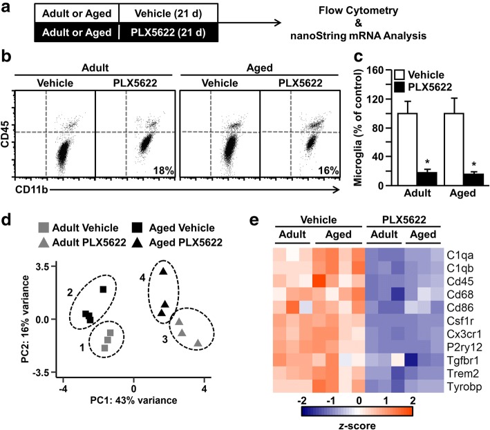
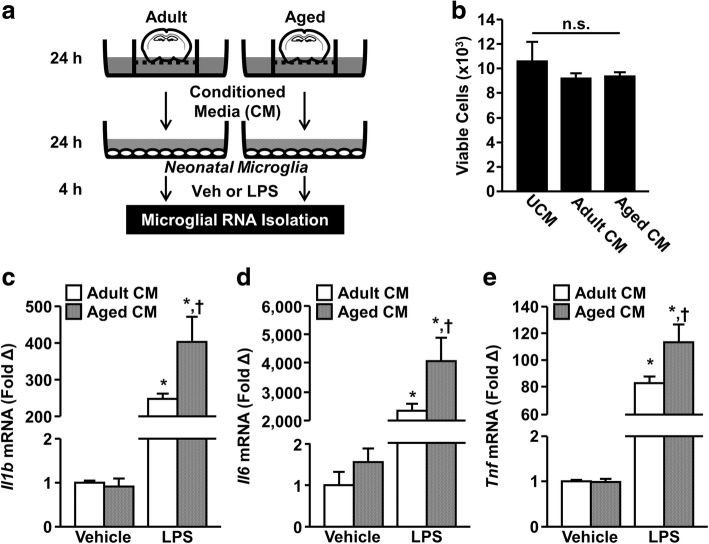
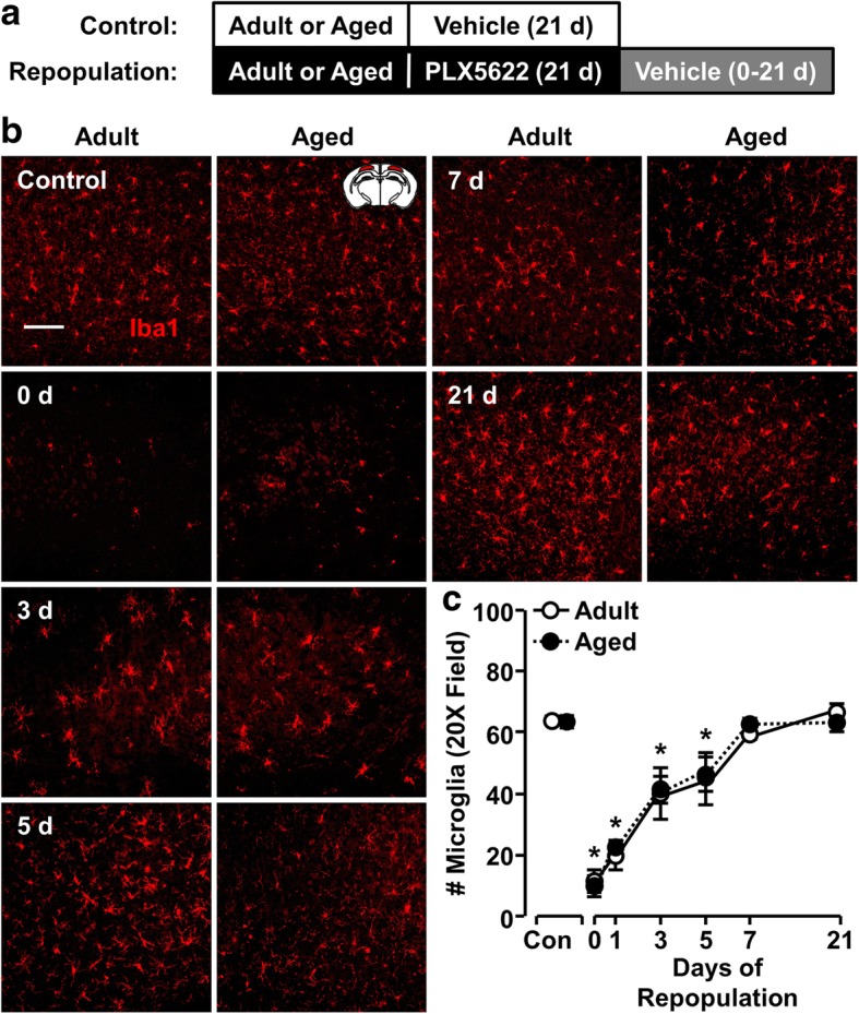
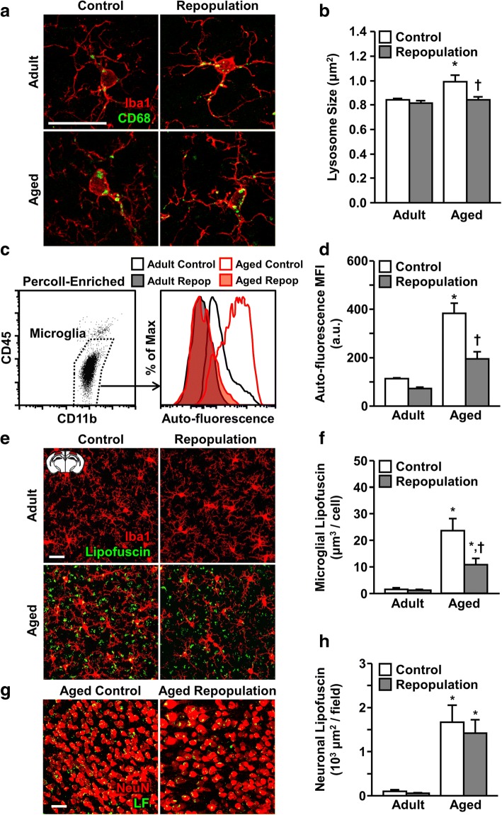
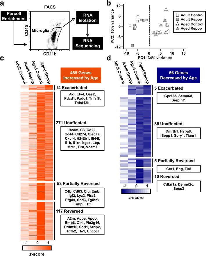
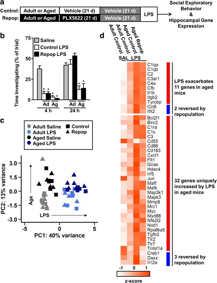
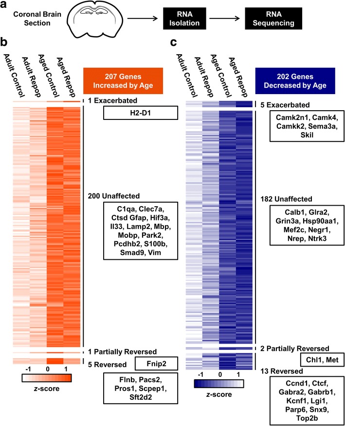
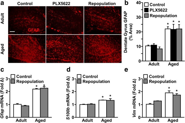
Microglia are the resident innate immune cells of the central nervous system. Limited turnover throughout the lifespan leaves microglia susceptible to age-associated dysfunction. Indeed, we and others have reported microglia develop a pro-inflammatory or "primed" profile with age, characterized by increased expression of inflammatory mediators (e.g., MHC-II, CD68, IL-1β). Moreover, immune challenge with lipopolysaccharide (LPS) causes an exaggerated and prolonged neuroinflammatory response mediated by primed microglia in the aged brain. Recent studies show colony-stimulating factor 1 receptor (CSF1R) antagonism results in rapid depletion of microglia without significant complications. Therefore, we hypothesized that CSF1R antagonist-mediated depletion of microglia in the aged brain would result in repopulation with new and unprimed microglia. Here we provide novel evidence that microglia in the brain of adult (6-8 weeks old) and aged (16-18 months old) BALB/c mice were depleted following 3-week oral PLX5622 administration. When CSF1R antagonism was stopped, microglia repopulated equally in the adult and aged brain. Microglial depletion and repopulation reversed age-associated increases in microglial CD68 lysosome enlargement and lipofuscin accumulation. Microglia-specific RNA sequencing revealed 511 differentially expressed genes with age. Of these, 117 genes were reversed by microglial repopulation (e.g., Apoe, Tgfb2, Socs3). Nevertheless, LPS challenge still induced an exaggerated microglial inflammatory response in the aged brain compared to adults. RNA sequencing of whole-brain tissue revealed an age-induced inflammatory signature, including reactive astrocytes, that was not restored by microglial depletion and repopulation. Furthermore, the microenvironment of the aged brain produced soluble factors that influenced developing microglia ex vivo and induced a profile primed to LPS challenge. Thus, the aged brain microenvironment promotes microglial priming despite repopulation of new microglia. Collectively, aged microglia proliferate and repopulate the brain, but these new cells still adopt a pro-inflammatory profile in the aged brain.
小胶质细胞是中枢神经系统的固有免疫细胞。在整个生命周期中,其更替有限,这使得小胶质细胞容易出现与年龄相关的功能障碍。事实上,我们和其他人已经报告说,小胶质细胞随着年龄的增长会发展出一种促炎或“预先激活”的表型,其特征是炎症介质(例如 MHC-II、CD68、IL-1β)的表达增加。此外,用脂多糖(LPS)进行免疫挑战会导致衰老大脑中预先激活的小胶质细胞引发过度和持久的神经炎症反应。最近的研究表明,集落刺激因子 1 受体(CSF1R)拮抗剂的使用会迅速耗尽小胶质细胞,而没有明显的并发症。因此,我们假设 CSF1R 拮抗剂介导的衰老大脑中小胶质细胞的耗竭会导致新的未预先激活的小胶质细胞的重新填充。在这里,我们提供了新的证据,表明在成年(6-8 周龄)和老年(16-18 月龄)BALB/c 小鼠的大脑中,在接受 3 周口服 PLX5622 治疗后,小胶质细胞被耗尽。当 CSF1R 拮抗作用停止时,小胶质细胞在成年和老年大脑中同样重新填充。小胶质细胞的耗竭和重新填充逆转了与年龄相关的小胶质细胞 CD68 溶酶体增大和脂褐素积累的增加。小胶质细胞特异性 RNA 测序显示,与年龄相关的差异表达基因有 511 个。其中,117 个基因被小胶质细胞的重新填充所逆转(例如,Apoe、Tgfb2、SocS3)。然而,与成年小鼠相比,LPS 挑战仍会在老年小鼠的大脑中引发过度的小胶质细胞炎症反应。对整个脑组织的 RNA 测序揭示了一个与年龄相关的炎症特征,包括反应性星形胶质细胞,而这些特征不会因小胶质细胞的耗竭和重新填充而恢复。此外,老年大脑的微环境产生了可溶性因子,这些因子会影响体外发育中的小胶质细胞,并诱导出一种对 LPS 挑战有预先激活作用的表型。因此,尽管新的小胶质细胞重新填充,但老年大脑的微环境仍会促进小胶质细胞的预先激活。总的来说,衰老的小胶质细胞会增殖并重新填充大脑,但这些新细胞在老年大脑中仍会呈现出促炎表型。