• 文献检索
  • 文档翻译
  • 深度研究
  • 学术资讯
  • Suppr Zotero 插件Zotero 插件
  • 邀请有礼
  • 套餐&价格
  • 历史记录
应用&插件
Suppr Zotero 插件Zotero 插件浏览器插件Mac 客户端Windows 客户端微信小程序
定价
高级版会员购买积分包购买API积分包
服务
文献检索文档翻译深度研究API 文档MCP 服务
关于我们
关于 Suppr公司介绍联系我们用户协议隐私条款
关注我们

Suppr 超能文献

核心技术专利:CN118964589B侵权必究
粤ICP备2023148730 号-1Suppr @ 2026

文献检索

告别复杂PubMed语法,用中文像聊天一样搜索,搜遍4000万医学文献。AI智能推荐,让科研检索更轻松。

立即免费搜索

文件翻译

保留排版,准确专业,支持PDF/Word/PPT等文件格式,支持 12+语言互译。

免费翻译文档

深度研究

AI帮你快速写综述,25分钟生成高质量综述,智能提取关键信息,辅助科研写作。

立即免费体验

分析连接组学的图像分割。

Analyzing Image Segmentation for Connectomics.

机构信息

Howard Hughes Medical Institute, Ashburn, VA, United States.

出版信息

Front Neural Circuits. 2018 Nov 13;12:102. doi: 10.3389/fncir.2018.00102. eCollection 2018.

DOI:10.3389/fncir.2018.00102
PMID:30483069
原文链接:https://pmc.ncbi.nlm.nih.gov/articles/PMC6243088/
Abstract

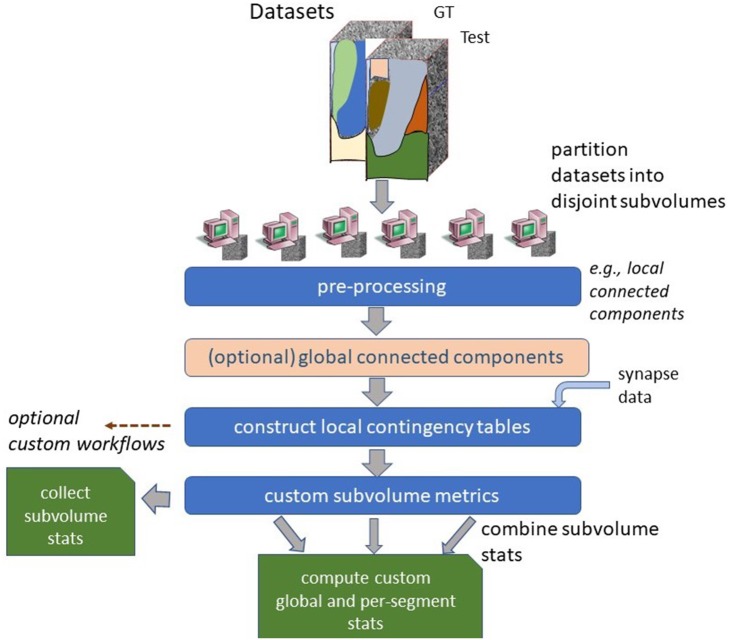
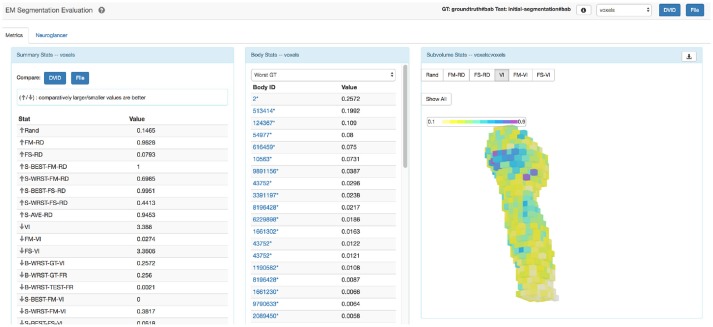
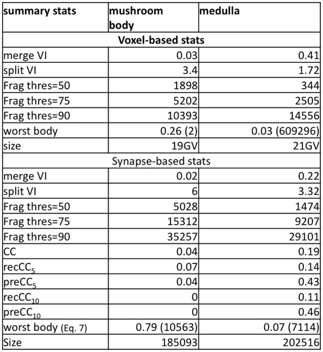
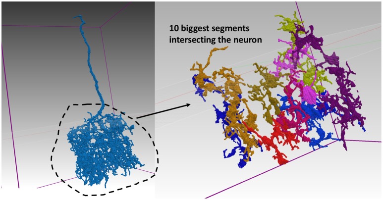
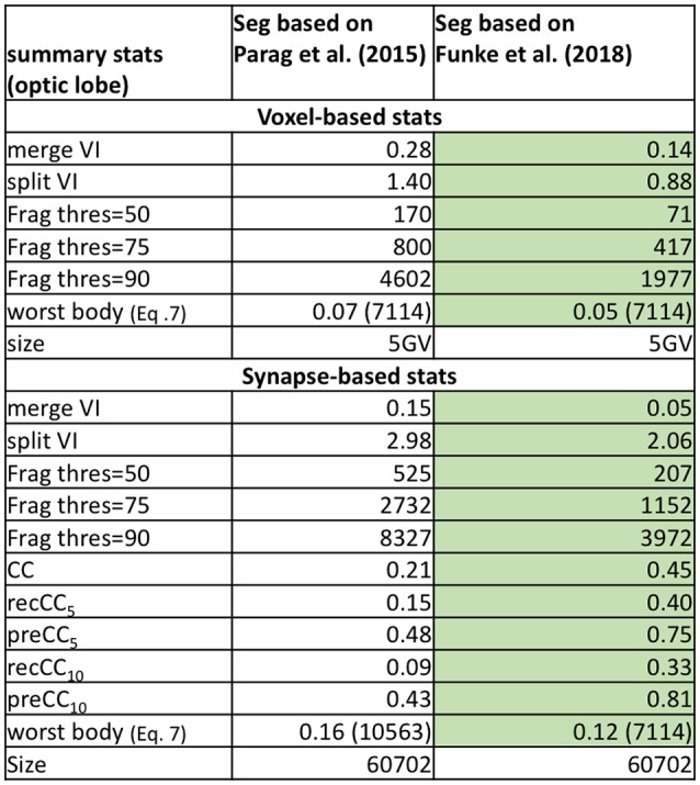
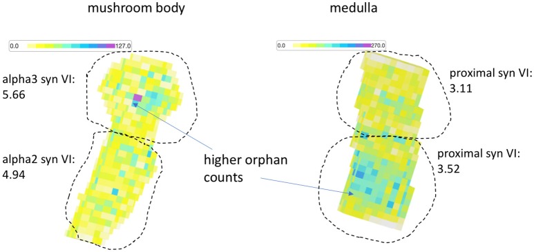
Automatic image segmentation is critical to scale up electron microscope (EM) connectome reconstruction. To this end, segmentation competitions, such as CREMI and SNEMI, exist to help researchers evaluate segmentation algorithms with the goal of improving them. Because generating ground truth is time-consuming, these competitions often fail to capture the challenges in segmenting larger datasets required in connectomics. More generally, the common metrics for EM image segmentation do not emphasize impact on downstream analysis and are often not very useful for isolating problem areas in the segmentation. For example, they do not capture connectivity information and often over-rate the quality of a segmentation as we demonstrate later. To address these issues, we introduce a novel strategy to enable evaluation of segmentation at large scales both in a supervised setting, where ground truth is available, or an unsupervised setting. To achieve this, we first introduce new metrics more closely aligned with the use of segmentation in downstream analysis and reconstruction. In particular, these include synapse connectivity and completeness metrics that provide both meaningful and intuitive interpretations of segmentation quality as it relates to the preservation of neuron connectivity. Also, we propose measures of segmentation correctness and completeness with respect to the percentage of "orphan" fragments and the concentrations of self-loops formed by segmentation failures, which are helpful in analysis and can be computed without ground truth. The introduction of new metrics intended to be used for practical applications involving large datasets necessitates a scalable software ecosystem, which is a critical contribution of this paper. To this end, we introduce a scalable, flexible software framework that enables integration of several different metrics and provides mechanisms to evaluate and debug differences between segmentations. We also introduce visualization software to help users to consume the various metrics collected. We evaluate our framework on two relatively large public groundtruth datasets providing novel insights on example segmentations.

摘要

自动图像分割对于扩大电子显微镜 (EM) 连接组重建至关重要。为此,存在分割竞赛,如 CREMI 和 SNEMI,以帮助研究人员评估分割算法,目的是改进它们。由于生成地面实况很耗时,这些竞赛往往无法捕捉到在连接组学中需要分割更大数据集的挑战。更一般地说,用于 EM 图像分割的常用指标不强调对下游分析的影响,并且通常对于隔离分割中的问题区域不是非常有用。例如,它们不捕获连接信息,并且经常像我们稍后演示的那样高估分割的质量。为了解决这些问题,我们引入了一种新策略,以便在有地面实况的监督设置或无监督设置下,在大规模上评估分割。为此,我们首先引入了与下游分析和重建中分割使用更紧密相关的新指标。特别是,这些指标包括突触连接性和完整性指标,它们提供了与神经元连接保持相关的分割质量的有意义和直观的解释。此外,我们提出了针对“孤儿”片段的百分比和由分割失败形成的自循环浓度的分割正确性和完整性度量,这些度量有助于分析并且可以在没有地面实况的情况下计算。引入旨在用于涉及大数据集的实际应用的新指标需要可扩展的软件生态系统,这是本文的一个关键贡献。为此,我们引入了一个可扩展的、灵活的软件框架,该框架能够集成几种不同的指标,并提供评估和调试分割之间差异的机制。我们还引入了可视化软件来帮助用户使用收集到的各种指标。我们在两个相对较大的公共地面实况数据集上评估了我们的框架,为示例分割提供了新的见解。

https://cdn.ncbi.nlm.nih.gov/pmc/blobs/b77e/6243088/17dd802629e5/fncir-12-00102-g0010.jpg
https://cdn.ncbi.nlm.nih.gov/pmc/blobs/b77e/6243088/00038b35489f/fncir-12-00102-g0001.jpg
https://cdn.ncbi.nlm.nih.gov/pmc/blobs/b77e/6243088/45bd99ad450b/fncir-12-00102-g0002.jpg
https://cdn.ncbi.nlm.nih.gov/pmc/blobs/b77e/6243088/2de1adb91714/fncir-12-00102-g0003.jpg
https://cdn.ncbi.nlm.nih.gov/pmc/blobs/b77e/6243088/ebc7f4665815/fncir-12-00102-g0004.jpg
https://cdn.ncbi.nlm.nih.gov/pmc/blobs/b77e/6243088/fa6db8affb94/fncir-12-00102-g0005.jpg
https://cdn.ncbi.nlm.nih.gov/pmc/blobs/b77e/6243088/31310a05f2b7/fncir-12-00102-g0006.jpg
https://cdn.ncbi.nlm.nih.gov/pmc/blobs/b77e/6243088/806eb2e404cb/fncir-12-00102-g0007.jpg
https://cdn.ncbi.nlm.nih.gov/pmc/blobs/b77e/6243088/c49605cd993a/fncir-12-00102-g0008.jpg
https://cdn.ncbi.nlm.nih.gov/pmc/blobs/b77e/6243088/3f939063257c/fncir-12-00102-g0009.jpg
https://cdn.ncbi.nlm.nih.gov/pmc/blobs/b77e/6243088/17dd802629e5/fncir-12-00102-g0010.jpg

相似文献

1
Analyzing Image Segmentation for Connectomics.分析连接组学的图像分割。
Front Neural Circuits. 2018 Nov 13;12:102. doi: 10.3389/fncir.2018.00102. eCollection 2018.
2
NeuTu: Software for Collaborative, Large-Scale, Segmentation-Based Connectome Reconstruction.NeuTu:用于协作式、大规模、基于分割的连接组重建的软件。
Front Neural Circuits. 2018 Nov 13;12:101. doi: 10.3389/fncir.2018.00101. eCollection 2018.
3
mEMbrain: an interactive deep learning MATLAB tool for connectomic segmentation on commodity desktops.mEMbrain:一个交互式深度学习 MATLAB 工具,用于在商用台式机上进行连接组分割。
Front Neural Circuits. 2023 Jun 15;17:952921. doi: 10.3389/fncir.2023.952921. eCollection 2023.
4
VAST (Volume Annotation and Segmentation Tool): Efficient Manual and Semi-Automatic Labeling of Large 3D Image Stacks.VAST(体积注释和分割工具):高效的大型 3D 图像堆栈手动和半自动标注。
Front Neural Circuits. 2018 Oct 16;12:88. doi: 10.3389/fncir.2018.00088. eCollection 2018.
5
Rapid and Semi-automated Extraction of Neuronal Cell Bodies and Nuclei from Electron Microscopy Image Stacks.从电子显微镜图像堆栈中快速半自动提取神经元细胞体和细胞核
Methods Mol Biol. 2016;1427:277-90. doi: 10.1007/978-1-4939-3615-1_16.
6
Fully-Automatic Synapse Prediction and Validation on a Large Data Set.全自动突触预测及其在大数据集上的验证。
Front Neural Circuits. 2018 Oct 29;12:87. doi: 10.3389/fncir.2018.00087. eCollection 2018.
7
LinSEM: Linearizing segmentation evaluation metrics for medical images.LinSEM:医学图像分割评估指标的线性化。
Med Image Anal. 2020 Feb;60:101601. doi: 10.1016/j.media.2019.101601. Epub 2019 Nov 9.
8
Unsupervised segmentation of noisy electron microscopy images using salient watersheds and region merging.基于显著分水岭和区域合并的无监督的含噪电子显微镜图像分割。
BMC Bioinformatics. 2013 Oct 4;14:294. doi: 10.1186/1471-2105-14-294.
9
A modular hierarchical approach to 3D electron microscopy image segmentation.一种用于三维电子显微镜图像分割的模块化分层方法。
J Neurosci Methods. 2014 Apr 15;226:88-102. doi: 10.1016/j.jneumeth.2014.01.022. Epub 2014 Jan 31.
10
Learning context cues for synapse segmentation.学习突触分割的上下文线索。
IEEE Trans Med Imaging. 2013 Oct;32(10):1864-77. doi: 10.1109/TMI.2013.2267747. Epub 2013 Jun 11.

引用本文的文献

1
Densely Populated Cell and Organelles Segmentation with U-Net Ensembles.基于U-Net集成的密集细胞和细胞器分割
bioRxiv. 2025 Jan 7:2024.11.19.623228. doi: 10.1101/2024.11.19.623228.
2
CEM500K, a large-scale heterogeneous unlabeled cellular electron microscopy image dataset for deep learning.CEM500K,一个用于深度学习的大规模异质无标签细胞电子显微镜图像数据集。
Elife. 2021 Apr 8;10:e65894. doi: 10.7554/eLife.65894.
3
Anatomy and activity patterns in a multifunctional motor neuron and its surrounding circuits.多功能运动神经元及其周围回路的解剖结构和活动模式。

本文引用的文献

1
DVID: Distributed Versioned Image-Oriented Dataservice.DVID:分布式版本化面向图像的数据服务。
Front Neural Circuits. 2019 Feb 5;13:5. doi: 10.3389/fncir.2019.00005. eCollection 2019.
2
Neural Reconstruction Integrity: A Metric for Assessing the Connectivity Accuracy of Reconstructed Neural Networks.神经重建完整性:一种评估重建神经网络连接准确性的指标。
Front Neuroinform. 2018 Nov 5;12:74. doi: 10.3389/fninf.2018.00074. eCollection 2018.
3
A Complete Electron Microscopy Volume of the Brain of Adult Drosophila melanogaster.
Elife. 2021 Feb 15;10:e61881. doi: 10.7554/eLife.61881.
4
Transforming the development and dissemination of cutting-edge microscopy and computation.推动前沿显微镜和计算技术的发展和传播。
Nat Methods. 2019 Aug;16(8):667-669. doi: 10.1038/s41592-019-0475-y.
成年黑腹果蝇大脑的完整电子显微镜体积。
Cell. 2018 Jul 26;174(3):730-743.e22. doi: 10.1016/j.cell.2018.06.019. Epub 2018 Jul 19.
4
High-precision automated reconstruction of neurons with flood-filling networks.基于填充网络的高精度自动化神经元重建。
Nat Methods. 2018 Aug;15(8):605-610. doi: 10.1038/s41592-018-0049-4. Epub 2018 Jul 16.
5
Large Scale Image Segmentation with Structured Loss Based Deep Learning for Connectome Reconstruction.基于深度学习的结构化损失的大规模图像分割在连接组重构中的应用。
IEEE Trans Pattern Anal Mach Intell. 2019 Jul;41(7):1669-1680. doi: 10.1109/TPAMI.2018.2835450. Epub 2018 May 24.
6
Conserved neural circuit structure across larval development revealed by comparative connectomics.通过比较连接组学揭示的幼虫发育过程中保守的神经回路结构。
Elife. 2017 Oct 23;6:e29089. doi: 10.7554/eLife.29089.
7
A connectome of a learning and memory center in the adult brain.成人大脑学习与记忆中心的连接组。
Elife. 2017 Jul 18;6:e26975. doi: 10.7554/eLife.26975.
8
TED: A Tolerant Edit Distance for segmentation evaluation.TED:用于分割评估的容忍编辑距离。
Methods. 2017 Feb 15;115:119-127. doi: 10.1016/j.ymeth.2016.12.013. Epub 2017 Jan 17.
9
Quantitative neuroanatomy for connectomics in Drosophila.果蝇连接组学的定量神经解剖学
Elife. 2016 Mar 18;5:e12059. doi: 10.7554/eLife.12059.
10
Crowdsourcing the creation of image segmentation algorithms for connectomics.众包创建用于连接组学的图像分割算法。
Front Neuroanat. 2015 Nov 5;9:142. doi: 10.3389/fnana.2015.00142. eCollection 2015.