• 文献检索
  • 文档翻译
  • 深度研究
  • 学术资讯
  • Suppr Zotero 插件Zotero 插件
  • 邀请有礼
  • 套餐&价格
  • 历史记录
应用&插件
Suppr Zotero 插件Zotero 插件浏览器插件Mac 客户端Windows 客户端微信小程序
定价
高级版会员购买积分包购买API积分包
服务
文献检索文档翻译深度研究API 文档MCP 服务
关于我们
关于 Suppr公司介绍联系我们用户协议隐私条款
关注我们

Suppr 超能文献

核心技术专利:CN118964589B侵权必究
粤ICP备2023148730 号-1Suppr @ 2026

文献检索

告别复杂PubMed语法,用中文像聊天一样搜索,搜遍4000万医学文献。AI智能推荐,让科研检索更轻松。

立即免费搜索

文件翻译

保留排版,准确专业,支持PDF/Word/PPT等文件格式,支持 12+语言互译。

免费翻译文档

深度研究

AI帮你快速写综述,25分钟生成高质量综述,智能提取关键信息,辅助科研写作。

立即免费体验

CEM500K,一个用于深度学习的大规模异质无标签细胞电子显微镜图像数据集。

CEM500K, a large-scale heterogeneous unlabeled cellular electron microscopy image dataset for deep learning.

机构信息

Center for Molecular Microscopy, Center for Cancer Research, National Cancer Institute, National Institutes of Health, Bethesda, United States.

Cancer Research Technology Program, Frederick National Laboratory for Cancer Research, Frederick, United States.

出版信息

Elife. 2021 Apr 8;10:e65894. doi: 10.7554/eLife.65894.

DOI:10.7554/eLife.65894
PMID:33830015
原文链接:https://pmc.ncbi.nlm.nih.gov/articles/PMC8032397/
Abstract

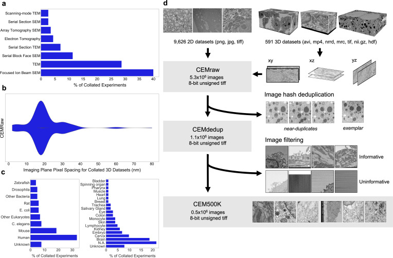
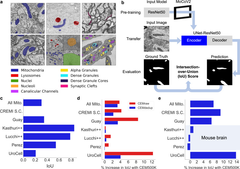
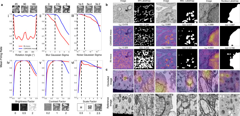
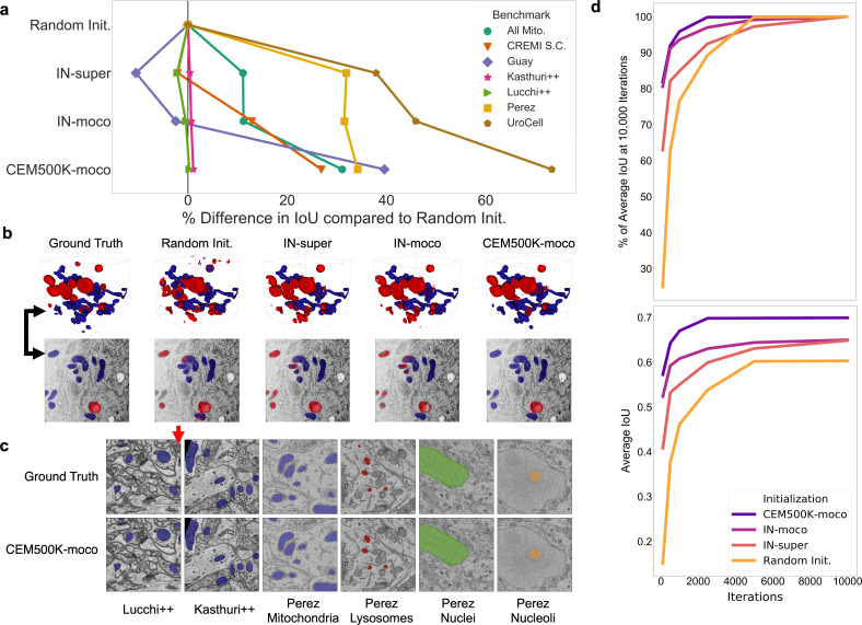
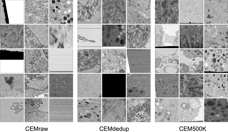
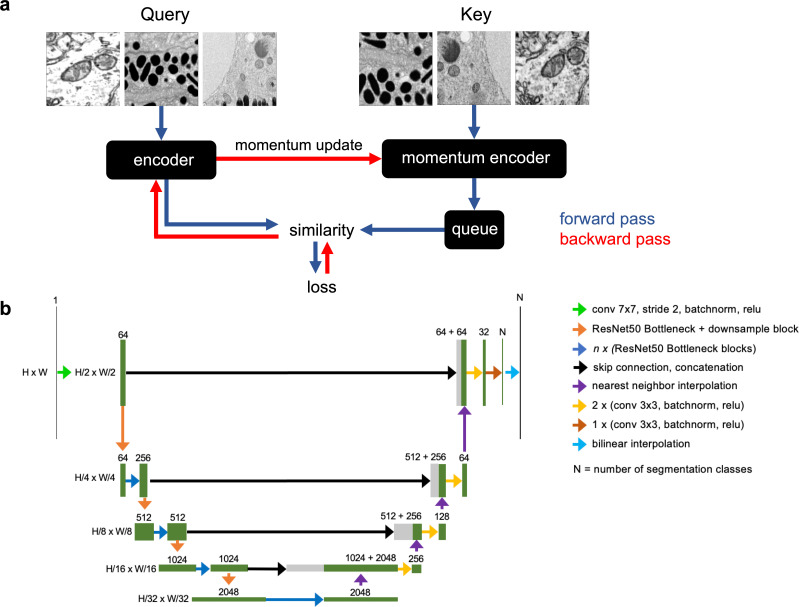
Automated segmentation of cellular electron microscopy (EM) datasets remains a challenge. Supervised deep learning (DL) methods that rely on region-of-interest (ROI) annotations yield models that fail to generalize to unrelated datasets. Newer unsupervised DL algorithms require relevant pre-training images, however, pre-training on currently available EM datasets is computationally expensive and shows little value for unseen biological contexts, as these datasets are large and homogeneous. To address this issue, we present CEM500K, a nimble 25 GB dataset of 0.5 × 10 unique 2D cellular EM images curated from nearly 600 three-dimensional (3D) and 10,000 two-dimensional (2D) images from >100 unrelated imaging projects. We show that models pre-trained on CEM500K learn features that are biologically relevant and resilient to meaningful image augmentations. Critically, we evaluate transfer learning from these pre-trained models on six publicly available and one newly derived benchmark segmentation task and report state-of-the-art results on each. We release the CEM500K dataset, pre-trained models and curation pipeline for model building and further expansion by the EM community. Data and code are available at https://www.ebi.ac.uk/pdbe/emdb/empiar/entry/10592/ and https://git.io/JLLTz.

摘要

细胞电子显微镜 (EM) 数据集的自动分割仍然是一个挑战。依赖于感兴趣区域 (ROI) 注释的监督深度学习 (DL) 方法生成的模型无法推广到不相关的数据集。较新的无监督 DL 算法需要相关的预训练图像,但是,在当前可用的 EM 数据集上进行预训练在计算上是昂贵的,并且对于看不见的生物背景几乎没有价值,因为这些数据集很大且同质。为了解决这个问题,我们提出了 CEM500K,这是一个灵活的 25GB 数据集,包含 0.5×10 个独特的 2D 细胞 EM 图像,这些图像是从近 600 个 3D 和 10000 个 2D 图像中提取出来的,这些图像来自于 >100 个不相关的成像项目。我们表明,在 CEM500K 上预训练的模型学习到的特征是具有生物学意义的,并且对有意义的图像增强具有弹性。至关重要的是,我们评估了这些预训练模型在六个公开可用的和一个新衍生的基准分割任务中的迁移学习,并在每个任务上报告了最先进的结果。我们发布了 CEM500K 数据集、预训练模型和模型构建的策展管道,以供 EM 社区进一步扩展。数据和代码可在 https://www.ebi.ac.uk/pdbe/emdb/empiar/entry/10592/ 和 https://git.io/JLLTz 获得。

https://cdn.ncbi.nlm.nih.gov/pmc/blobs/f4ec/8032397/551c3f96bb13/elife-65894-app1-fig7.jpg
https://cdn.ncbi.nlm.nih.gov/pmc/blobs/f4ec/8032397/aa7095b2ec0d/elife-65894-fig1.jpg
https://cdn.ncbi.nlm.nih.gov/pmc/blobs/f4ec/8032397/85a783d96132/elife-65894-fig2.jpg
https://cdn.ncbi.nlm.nih.gov/pmc/blobs/f4ec/8032397/32689c15df64/elife-65894-fig3.jpg
https://cdn.ncbi.nlm.nih.gov/pmc/blobs/f4ec/8032397/4d6e0b221738/elife-65894-fig4.jpg
https://cdn.ncbi.nlm.nih.gov/pmc/blobs/f4ec/8032397/7a5070106b15/elife-65894-app1-fig1.jpg
https://cdn.ncbi.nlm.nih.gov/pmc/blobs/f4ec/8032397/f43db4461547/elife-65894-app1-fig2.jpg
https://cdn.ncbi.nlm.nih.gov/pmc/blobs/f4ec/8032397/06fbafa57044/elife-65894-app1-fig3.jpg
https://cdn.ncbi.nlm.nih.gov/pmc/blobs/f4ec/8032397/e5f24cd473a3/elife-65894-app1-fig4.jpg
https://cdn.ncbi.nlm.nih.gov/pmc/blobs/f4ec/8032397/c1fcf4e4d3db/elife-65894-app1-fig5.jpg
https://cdn.ncbi.nlm.nih.gov/pmc/blobs/f4ec/8032397/6cb6153ef479/elife-65894-app1-fig6.jpg
https://cdn.ncbi.nlm.nih.gov/pmc/blobs/f4ec/8032397/551c3f96bb13/elife-65894-app1-fig7.jpg

相似文献

1
CEM500K, a large-scale heterogeneous unlabeled cellular electron microscopy image dataset for deep learning.CEM500K,一个用于深度学习的大规模异质无标签细胞电子显微镜图像数据集。
Elife. 2021 Apr 8;10:e65894. doi: 10.7554/eLife.65894.
2
Segmentation in large-scale cellular electron microscopy with deep learning: A literature survey.基于深度学习的大规模细胞电子显微镜图像分割:文献综述。
Med Image Anal. 2023 Oct;89:102920. doi: 10.1016/j.media.2023.102920. Epub 2023 Aug 6.
3
Deep learning based domain adaptation for mitochondria segmentation on EM volumes.基于深度学习的 EM 体数据中线粒体分割领域自适应方法
Comput Methods Programs Biomed. 2022 Jul;222:106949. doi: 10.1016/j.cmpb.2022.106949. Epub 2022 Jun 14.
4
Instance segmentation of mitochondria in electron microscopy images with a generalist deep learning model trained on a diverse dataset.使用在多样化数据集上训练的通用深度学习模型对电子显微镜图像中的线粒体进行实例分割。
Cell Syst. 2023 Jan 18;14(1):58-71.e5. doi: 10.1016/j.cels.2022.12.006.
5
Bi-channel image registration and deep-learning segmentation (BIRDS) for efficient, versatile 3D mapping of mouse brain.双通道图像配准和深度学习分割(BIRDS)用于高效、通用的小鼠大脑 3D 映射。
Elife. 2021 Jan 18;10:e63455. doi: 10.7554/eLife.63455.
6
Multiscale unsupervised domain adaptation for automatic pancreas segmentation in CT volumes using adversarial learning.基于对抗学习的 CT 容积中多尺度无监督域自适应自动胰腺分割。
Med Phys. 2022 Sep;49(9):5799-5818. doi: 10.1002/mp.15827. Epub 2022 Jul 27.
7
Local contrastive loss with pseudo-label based self-training for semi-supervised medical image segmentation.基于伪标签自训练的局部对比损失的半监督医学图像分割。
Med Image Anal. 2023 Jul;87:102792. doi: 10.1016/j.media.2023.102792. Epub 2023 Mar 11.
8
Generalizing Deep Learning for Medical Image Segmentation to Unseen Domains via Deep Stacked Transformation.通过深度堆叠变换将深度学习用于医学图像分割推广到未见领域。
IEEE Trans Med Imaging. 2020 Jul;39(7):2531-2540. doi: 10.1109/TMI.2020.2973595. Epub 2020 Feb 12.
9
On the objectivity, reliability, and validity of deep learning enabled bioimage analyses.深度学习赋能的生物影像分析的客观性、可靠性和有效性。
Elife. 2020 Oct 19;9:e59780. doi: 10.7554/eLife.59780.
10
TEM virus images: Benchmark dataset and deep learning classification.TEM 病毒图像:基准数据集和深度学习分类。
Comput Methods Programs Biomed. 2021 Sep;209:106318. doi: 10.1016/j.cmpb.2021.106318. Epub 2021 Jul 29.

引用本文的文献

1
MIFA: Metadata, Incentives, Formats and Accessibility guidelines to improve the reuse of AI datasets for bioimage analysis.MIFA:用于改善生物图像分析中人工智能数据集再利用的元数据、激励措施、格式和可访问性指南。
Nat Methods. 2025 Sep 15. doi: 10.1038/s41592-025-02835-8.
2
DeepEM Playground: Bringing deep learning to electron microscopy labs.深度电子显微镜演练场:将深度学习引入电子显微镜实验室。
J Microsc. 2025 Sep;299(3):287-300. doi: 10.1111/jmi.70005. Epub 2025 Jun 28.
3
Deep learning-driven automated mitochondrial segmentation for analysis of complex transmission electron microscopy images.

本文引用的文献

1
Deep learning for automatic segmentation of the nuclear envelope in electron microscopy data, trained with volunteer segmentations.利用志愿者分割标注进行训练的深度学习方法,用于自动分割电子显微镜数据中的核膜。
Traffic. 2021 Jul;22(7):240-253. doi: 10.1111/tra.12789. Epub 2021 May 16.
2
Dense cellular segmentation for EM using 2D-3D neural network ensembles.使用 2D-3D 神经网络集成进行 EM 的密集细胞分割。
Sci Rep. 2021 Jan 28;11(1):2561. doi: 10.1038/s41598-021-81590-0.
3
Semantic segmentation of HeLa cells: An objective comparison between one traditional algorithm and four deep-learning architectures.
深度学习驱动的自动线粒体分割,用于复杂透射电子显微镜图像分析。
Sci Rep. 2025 May 30;15(1):19076. doi: 10.1038/s41598-025-03311-1.
4
RETINA: Reconstruction-based pre-trained enhanced TransUNet for electron microscopy segmentation on the CEM500K dataset.视网膜:基于重建的预训练增强型TransUNet,用于CEM500K数据集上的电子显微镜图像分割
PLoS Comput Biol. 2025 May 28;21(5):e1013115. doi: 10.1371/journal.pcbi.1013115. eCollection 2025 May.
5
Volumetric Semantic Instance Segmentation of the Plasma Membrane of HeLa Cells.HeLa细胞质膜的体积语义实例分割
J Imaging. 2021 Jun 1;7(6):93. doi: 10.3390/jimaging7060093.
6
Modular segmentation, spatial analysis and visualization of volume electron microscopy datasets.体式电子显微镜数据集的模块化分割、空间分析和可视化。
Nat Protoc. 2024 May;19(5):1436-1466. doi: 10.1038/s41596-024-00957-5. Epub 2024 Feb 29.
7
Electron Microscopy Techniques for 3D Plant ER Imaging.用于植物内质网 3D 成像的电子显微镜技术。
Methods Mol Biol. 2024;2772:15-25. doi: 10.1007/978-1-0716-3710-4_2.
8
Morphomics via next-generation electron microscopy.基于下一代电子显微镜的形态计量学。
J Mol Cell Biol. 2024 Apr 10;15(12). doi: 10.1093/jmcb/mjad081.
9
Current Progress and Challenges in Large-Scale 3D Mitochondria Instance Segmentation.大规模 3D 线粒体实例分割的研究进展与挑战。
IEEE Trans Med Imaging. 2023 Dec;42(12):3956-3971. doi: 10.1109/TMI.2023.3320497. Epub 2023 Nov 30.
10
Volume electron microscopy.体积电子显微镜术
Nat Rev Methods Primers. 2022 Jul 7;2:51. doi: 10.1038/s43586-022-00131-9.
HeLa 细胞的语义分割:一种传统算法与四种深度学习架构的客观比较。
PLoS One. 2020 Oct 2;15(10):e0230605. doi: 10.1371/journal.pone.0230605. eCollection 2020.
4
Automatic segmentation of mitochondria and endolysosomes in volumetric electron microscopy data.在体积电子显微镜数据中对线粒体和内溶酶体进行自动分割。
Comput Biol Med. 2020 Apr;119:103693. doi: 10.1016/j.compbiomed.2020.103693. Epub 2020 Mar 3.
5
Correlative three-dimensional super-resolution and block-face electron microscopy of whole vitreously frozen cells.整体玻璃体冷冻细胞的相关三维超分辨率和块面电子显微镜。
Science. 2020 Jan 17;367(6475). doi: 10.1126/science.aaz5357.
6
The Relative Performance of Ensemble Methods with Deep Convolutional Neural Networks for Image Classification.深度卷积神经网络集成方法在图像分类中的相对性能
J Appl Stat. 2018;45(15):2800-2818. doi: 10.1080/02664763.2018.1441383. Epub 2018 Feb 26.
7
Quantitative 3D Mapping of the Human Skeletal Muscle Mitochondrial Network.人体骨骼肌线粒体网络的定量 3D 绘图。
Cell Rep. 2019 Jan 22;26(4):996-1009.e4. doi: 10.1016/j.celrep.2019.01.010. Epub 2019 Jan 15.
8
Detection of herpesvirus capsids in transmission electron microscopy images using transfer learning.利用迁移学习在透射电子显微镜图像中检测疱疹病毒衣壳
Histochem Cell Biol. 2019 Feb;151(2):101-114. doi: 10.1007/s00418-018-1759-5. Epub 2018 Nov 28.
9
Analyzing Image Segmentation for Connectomics.分析连接组学的图像分割。
Front Neural Circuits. 2018 Nov 13;12:102. doi: 10.3389/fncir.2018.00102. eCollection 2018.
10
A community-developed open-source computational ecosystem for big neuro data.一个由社区开发的用于处理大规模神经数据的开源计算生态系统。
Nat Methods. 2018 Nov;15(11):846-847. doi: 10.1038/s41592-018-0181-1.