Chaturvedi Abhishek, Rajiah Prabhakar, Croake Alexender, Saboo Sachin, Chaturvedi Apeksha
Imaging Science, University of Rochester Medical Center, 601, Elmwood Avenue, Rochester, NY, 14642, USA.
Radiology, University of Texas Southwestern Medical Center, Dallas, TX, USA.
Insights Imaging. 2018 Dec;9(6):989-1005. doi: 10.1007/s13244-018-0670-x. Epub 2018 Nov 27.
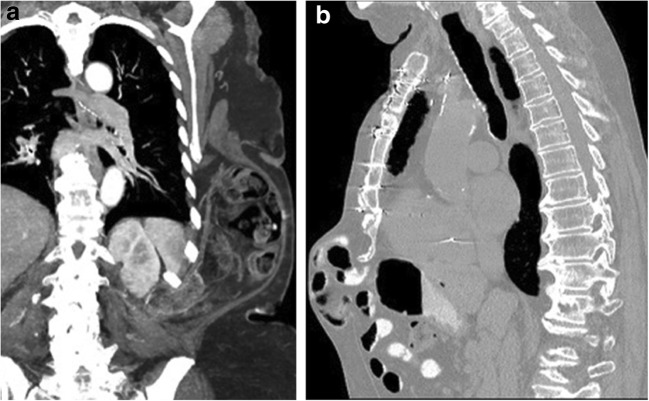
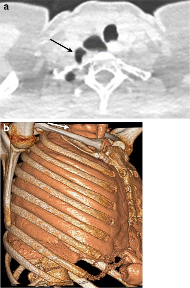
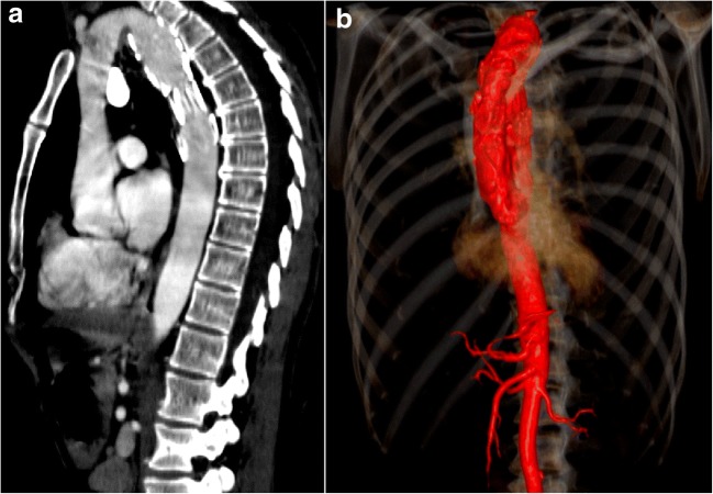
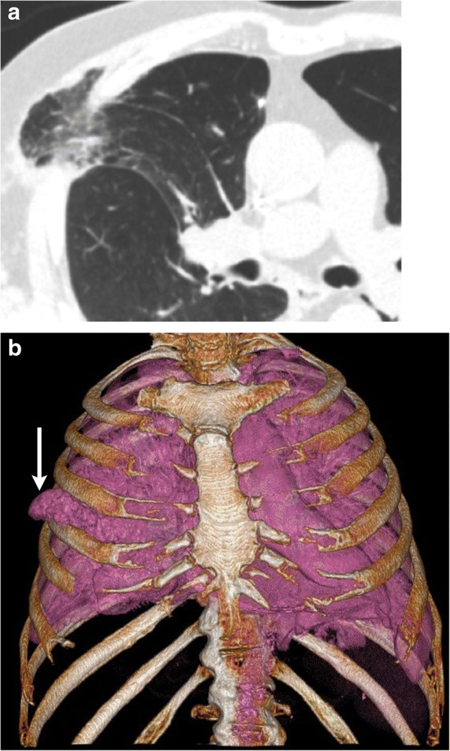
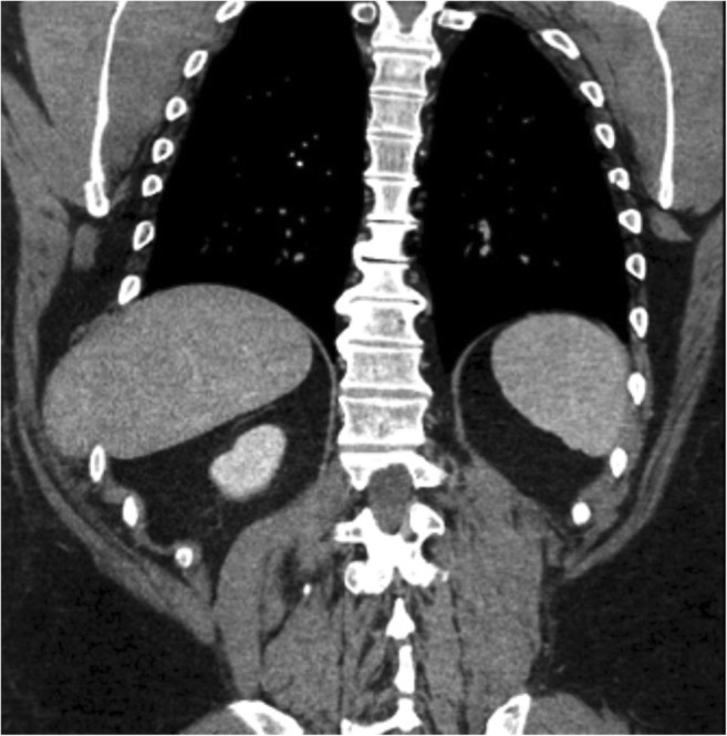
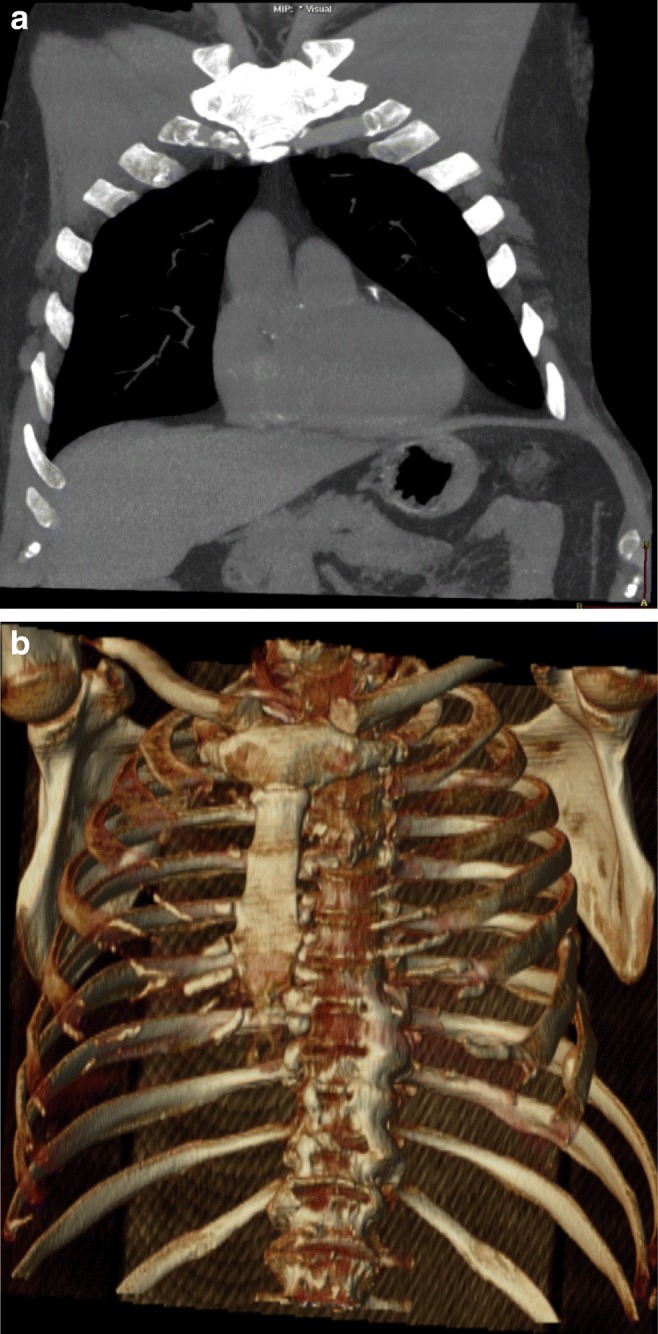
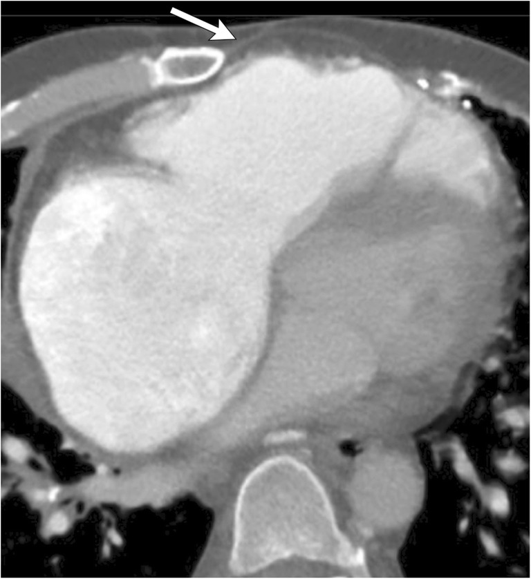
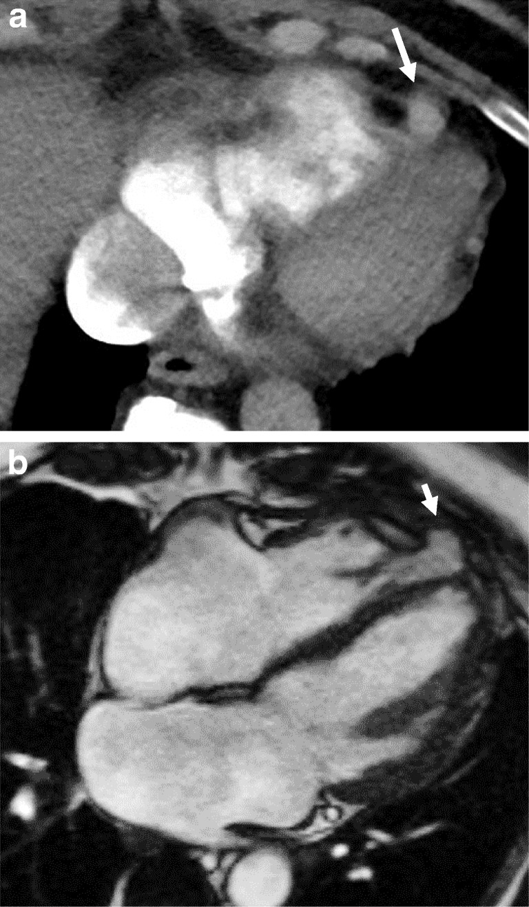
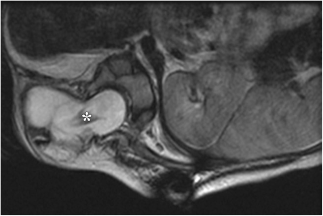
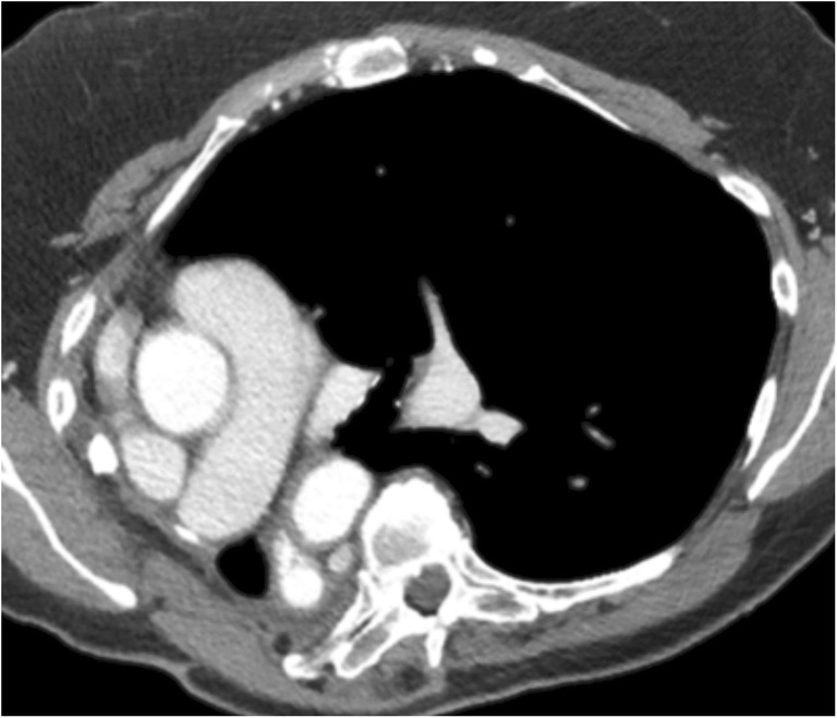
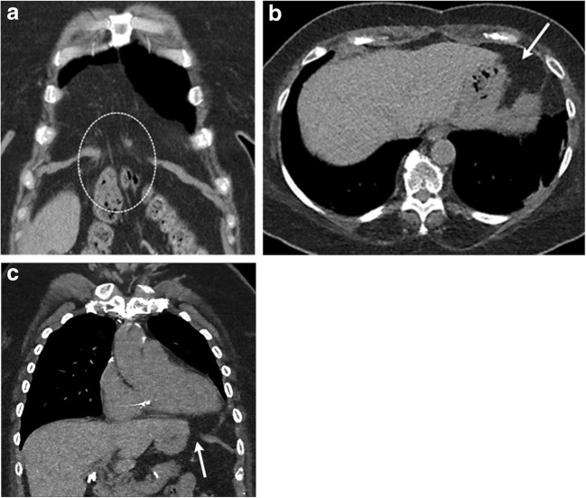
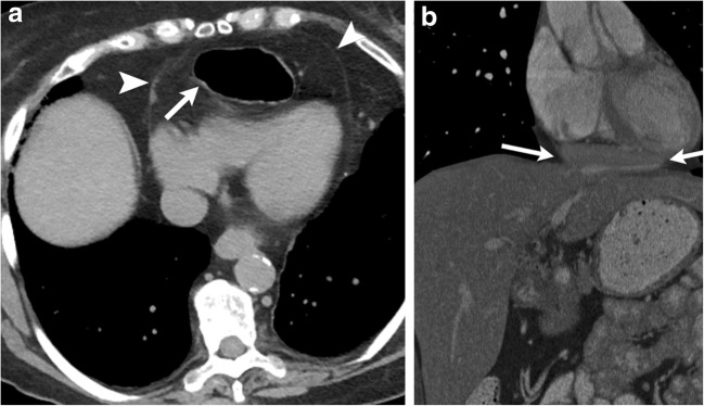
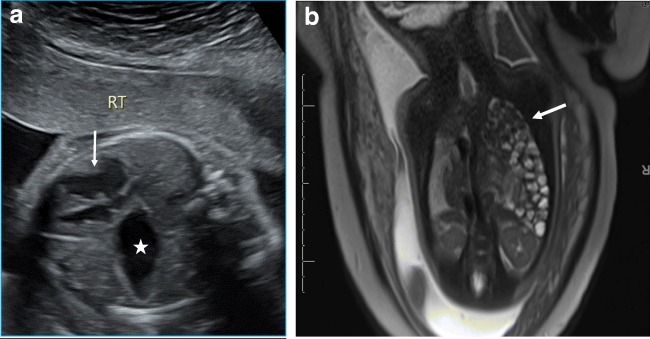
Thoracic hernias are characterised by either protrusion of the thoracic contents outside their normal anatomical confines or extension of the abdominal contents within the thorax. Thoracic hernias can be either congenital or acquired in aetiology. They can occur at the level of the thoracic inlet, chest wall or diaphragm. Thoracic hernias can be symptomatic or fortuitously discovered on imaging obtained for other indications. Complications of thoracic hernias include incarceration, trauma and strangulation with necrosis. Multiple imaging modalities are available to evaluate thoracic hernias. Radiographs usually offer the first clue to the diagnosis. Upper gastrointestinal radiography can identify bowel herniation and associated complications. CT and occasionally MR can be useful for further evaluation of these abnormalities, accurately identifying the type of hernia, its contents, associated complications, and provide a roadmap for surgical planning. In this article, we review the different types of thoracic hernias and the role of imaging in the evaluation of these hernias. TEACHING POINTS: • Protrusion of lung contents beyond the anatomic confines of the thorax constitutes a hernia. • Complications of thoracic hernias include incarceration, trauma and strangulation with necrosis. • Multiple imaging modalities are available to evaluate thoracic hernias. • CT is the imaging modality of choice for identifying thoracic hernias and their complications. • Imaging can provide a roadmap for surgical planning.
胸段疝的特征是胸腔内容物突出到其正常解剖边界之外,或者腹腔内容物延伸至胸腔内。胸段疝在病因上可分为先天性或后天性。它们可发生于胸廓入口、胸壁或膈肌水平。胸段疝可能有症状,也可能在因其他指征进行影像学检查时偶然发现。胸段疝的并发症包括嵌顿、创伤以及绞窄伴坏死。有多种成像方式可用于评估胸段疝。X线平片通常是诊断的首要线索。上消化道造影可识别肠疝及相关并发症。CT以及偶尔使用的MR对这些异常的进一步评估可能有用,可准确识别疝的类型、内容物、相关并发症,并为手术规划提供路线图。在本文中,我们回顾了不同类型的胸段疝以及成像在这些疝评估中的作用。教学要点:• 肺内容物突出到胸廓的解剖边界之外即构成疝。• 胸段疝的并发症包括嵌顿、创伤以及绞窄伴坏死。• 有多种成像方式可用于评估胸段疝。• CT是识别胸段疝及其并发症的首选成像方式。• 成像可为手术规划提供路线图。