Kushchayeva Yevgeniya S, Kushchayev Sergiy V, Glushko Tetiana Y, Tella Sri Harsha, Teytelboym Oleg M, Collins Michael T, Boyce Alison M
Diabetes, Endocrinology, and Obesity Branch, National Institute of Diabetes and Digestive and Kidney Diseases, National Institutes of Health, 31 Center Dr, Bethesda, MD, 20892, USA.
Division of Neuroradiology, Department of Radiology, Johns Hopkins Hospital, 1800 Orleans St, Baltimore, MD, 21287, USA.
Insights Imaging. 2018 Dec;9(6):1035-1056. doi: 10.1007/s13244-018-0666-6. Epub 2018 Nov 27.
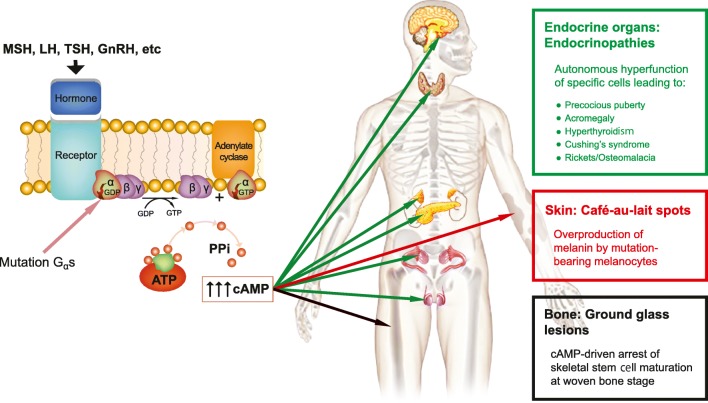
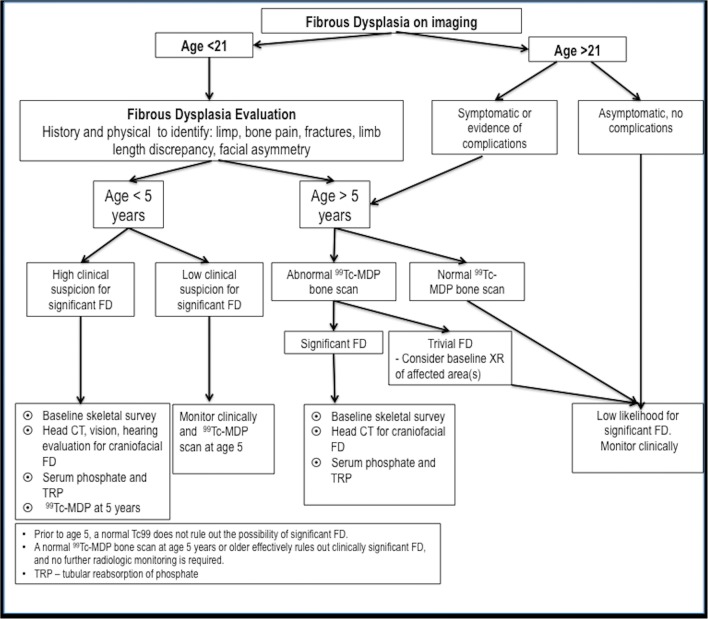
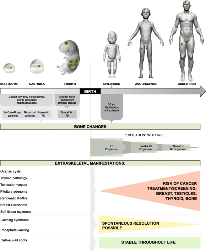
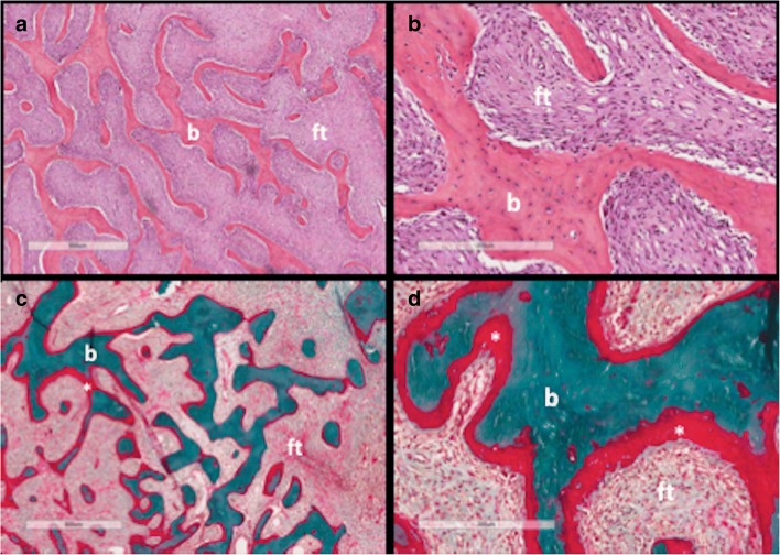
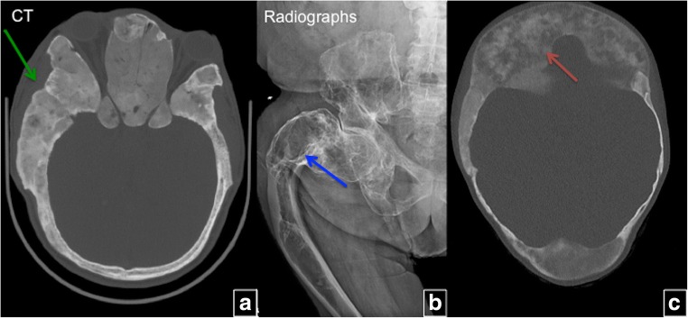
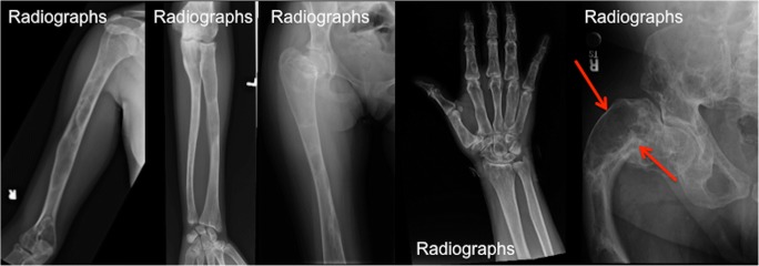
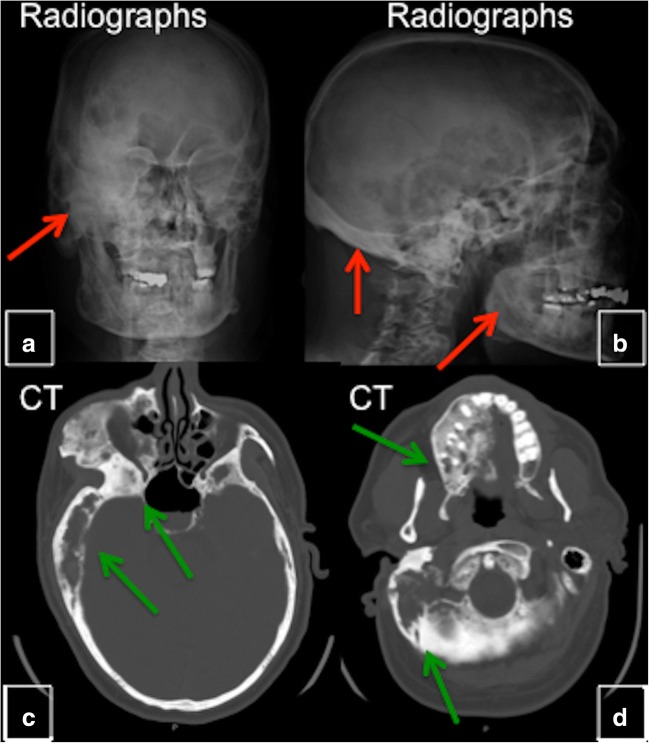
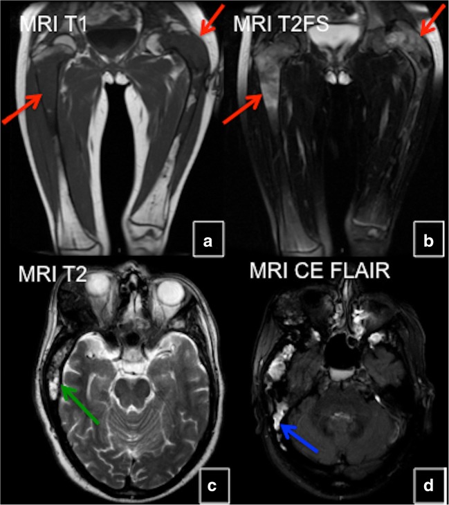
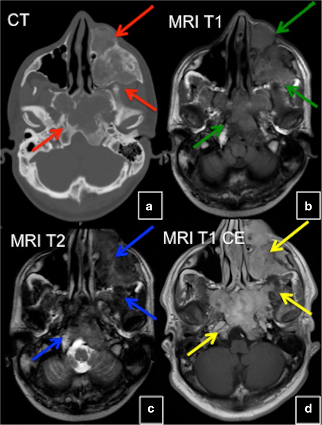
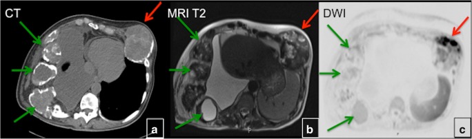
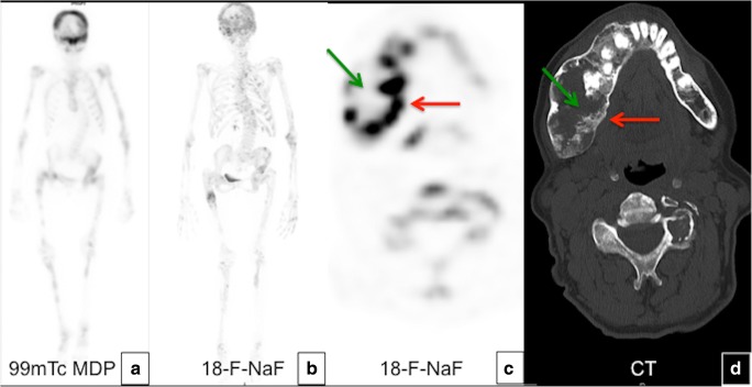
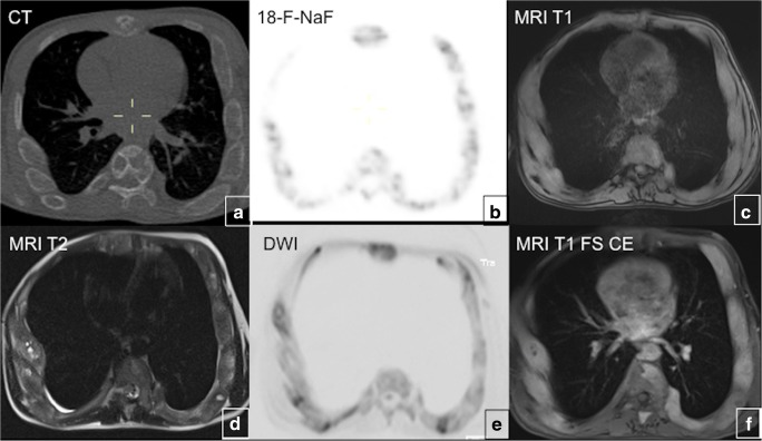
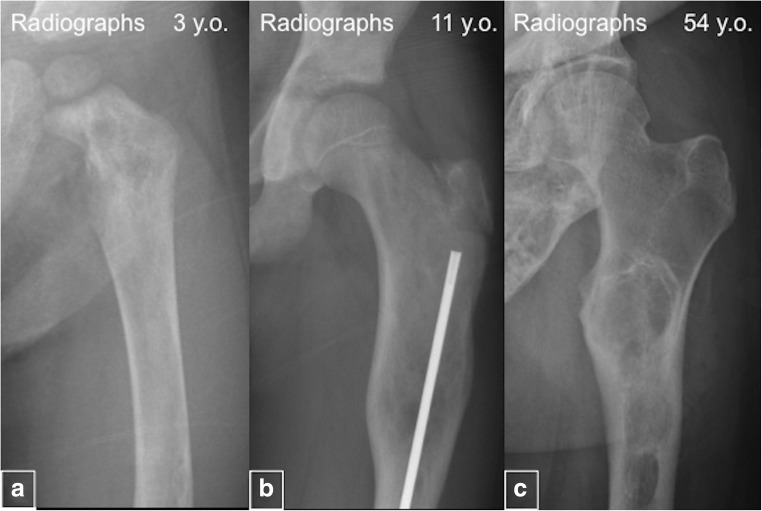
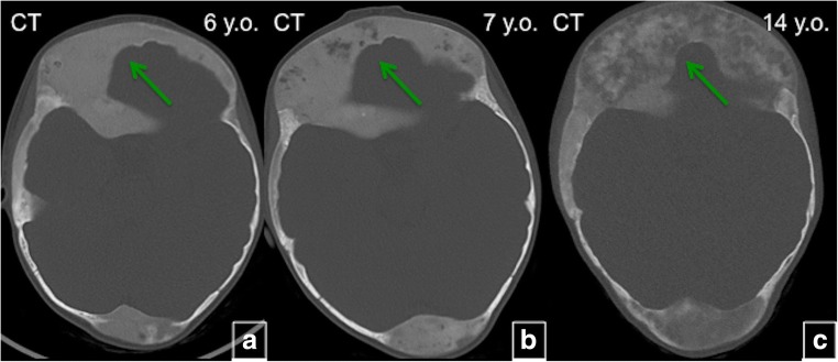
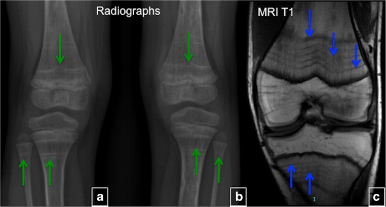
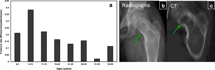
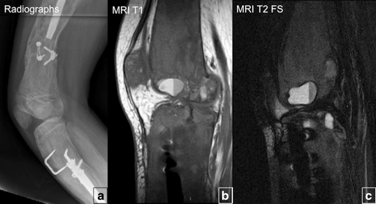
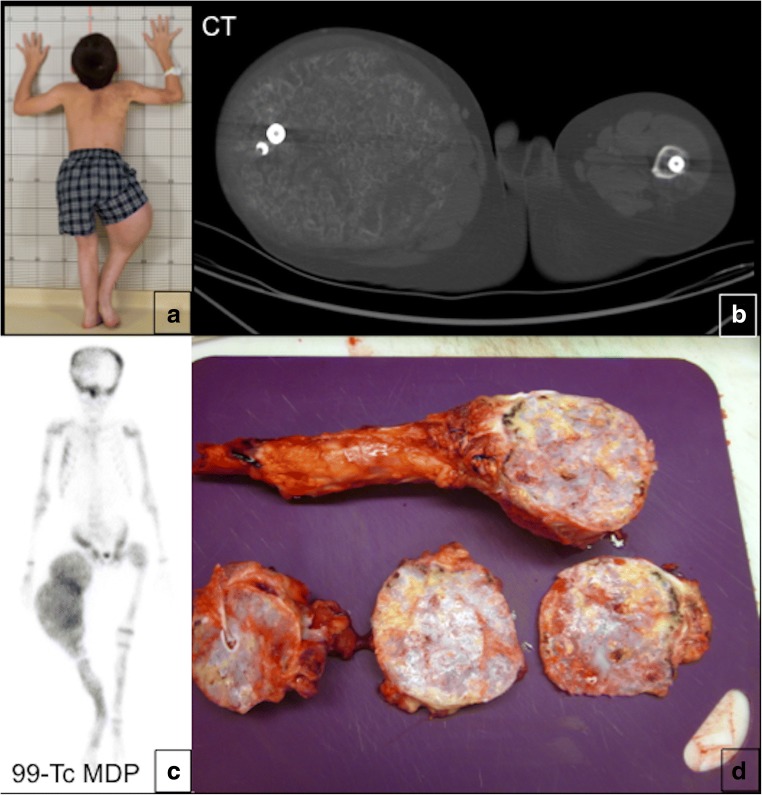
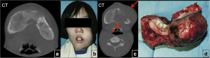
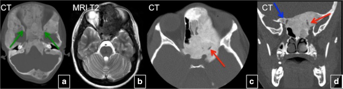
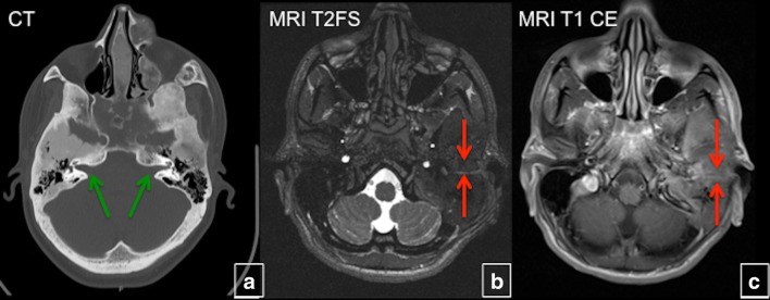
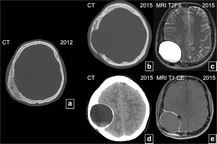
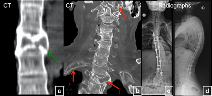
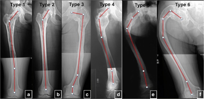
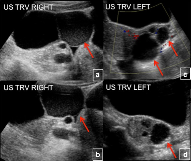
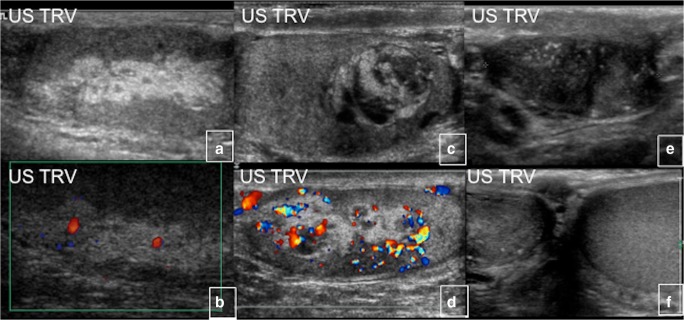
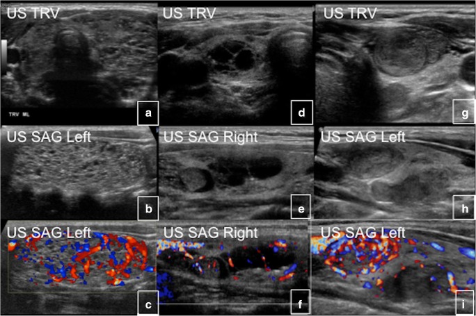
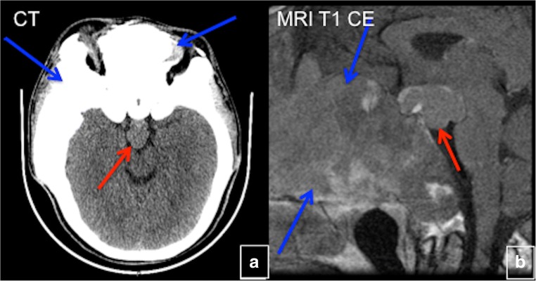
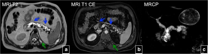
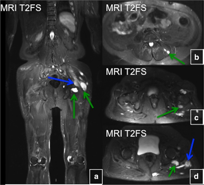
Fibrous dysplasia (FD) is a congenital disorder arising from sporadic mutation of the α-subunit of the Gs stimulatory protein. Osseous changes are characterised by the replacement and distortion of normal bone with poorly organised, structurally unsound, fibrous tissue. The disease process may be localised to a single or multiple bones. In McCune-Albright syndrome (MAS), fibrous dysplasia is associated with hyperfunction of endocrine organs and overproduction of melanin in the skin, while Mazabraud syndrome FD is associated with intramuscular myxomas. In radiology, FD is very often automatically associated with the term "ground glass matrix". However, FD is a complex disease, and knowledge of its unique pathogenesis and course are crucial to understanding imaging findings and potential complications. This article aims to not only summarise the spectrum of radiological findings of osseous and extra-osseous abnormalities associated with FD but also to highlight the pathological base of the disease evolution, corresponding imaging changes and complications based on the disease distribution. We also have provided current recommendations for clinical management and follow-up of patients with FD. TEACHING POINTS: • FD is often a part of complex disease, involving not only bone but also multiple other organs. • FD lesions are characterised by age-related histological, radiographical and clinical transformations. • Radiologists play a crucial role in the identification of osseous complications associated with FD. • The craniofacial form of the disease is the most common type of FD and the most difficult form to manage. • Patients with McCune-Albright syndrome may have different extra-skeletal abnormalities, which often require follow-up.
骨纤维异常增殖症(FD)是一种由Gs刺激蛋白α亚基的散发性突变引起的先天性疾病。骨质改变的特征是正常骨被结构紊乱、组织不良的纤维组织替代和变形。疾病过程可能局限于单骨或多骨。在McCune-Albright综合征(MAS)中,骨纤维异常增殖症与内分泌器官功能亢进及皮肤黑色素过度生成有关,而Mazabraud综合征中的骨纤维异常增殖症与肌内黏液瘤有关。在放射学中,骨纤维异常增殖症常常自动与术语“磨玻璃基质”联系在一起。然而,骨纤维异常增殖症是一种复杂的疾病,了解其独特的发病机制和病程对于理解影像学表现及潜在并发症至关重要。本文旨在不仅总结与骨纤维异常增殖症相关的骨内和骨外异常的放射学表现谱,还强调基于疾病分布的疾病演变的病理基础、相应的影像学变化及并发症。我们还提供了目前针对骨纤维异常增殖症患者临床管理和随访的建议。教学要点:•骨纤维异常增殖症通常是复杂疾病的一部分,不仅累及骨骼,还涉及多个其他器官。•骨纤维异常增殖症病变具有与年龄相关的组织学、影像学和临床转变特征。•放射科医生在识别与骨纤维异常增殖症相关的骨并发症方面起着关键作用。•疾病的颅面型是骨纤维异常增殖症最常见的类型,也是最难处理的类型。•McCune-Albright综合征患者可能有不同的骨骼外异常,常需要随访。