Department of Pathology, University of Cambridge, Cambridge, UK.
Department of Physiology, Development and Neuroscience, University of Cambridge, Cambridge, UK.
Nature. 2018 Dec;564(7735):263-267. doi: 10.1038/s41586-018-0753-3. Epub 2018 Nov 28.
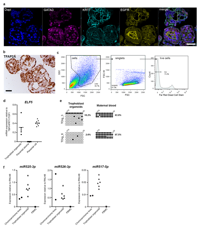
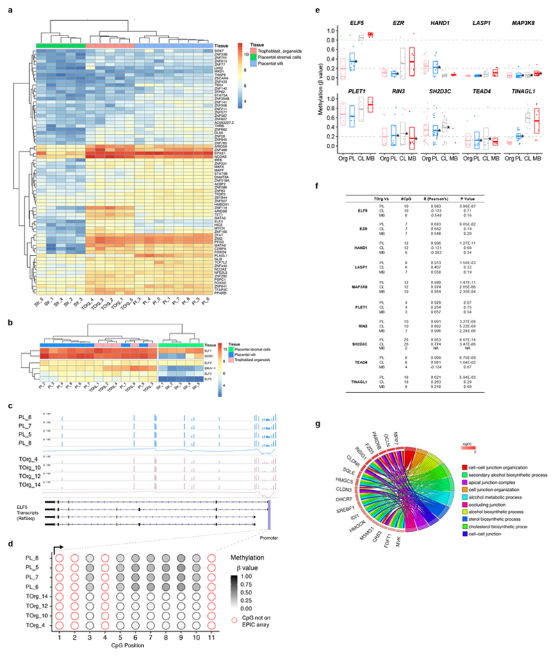
The placenta is the extraembryonic organ that supports the fetus during intrauterine life. Although placental dysfunction results in major disorders of pregnancy with immediate and lifelong consequences for the mother and child, our knowledge of the human placenta is limited owing to a lack of functional experimental models. After implantation, the trophectoderm of the blastocyst rapidly proliferates and generates the trophoblast, the unique cell type of the placenta. In vivo, proliferative villous cytotrophoblast cells differentiate into two main sub-populations: syncytiotrophoblast, the multinucleated epithelium of the villi responsible for nutrient exchange and hormone production, and extravillous trophoblast cells, which anchor the placenta to the maternal decidua and transform the maternal spiral arteries. Here we describe the generation of long-term, genetically stable organoid cultures of trophoblast that can differentiate into both syncytiotrophoblast and extravillous trophoblast. We used human leukocyte antigen (HLA) typing to confirm that the organoids were derived from the fetus, and verified their identities against four trophoblast-specific criteria. The cultures organize into villous-like structures, and we detected the secretion of placental-specific peptides and hormones, including human chorionic gonadotropin (hCG), growth differentiation factor 15 (GDF15) and pregnancy-specific glycoprotein (PSG) by mass spectrometry. The organoids also differentiate into HLA-G extravillous trophoblast cells, which vigorously invade in three-dimensional cultures. Analysis of the methylome reveals that the organoids closely resemble normal first trimester placentas. This organoid model will be transformative for studying human placental development and for investigating trophoblast interactions with the local and systemic maternal environment.
胎盘是支持胎儿宫内生命的胚胎外器官。尽管胎盘功能障碍导致妊娠的主要疾病,并对母婴产生即时和终身影响,但由于缺乏功能实验模型,我们对人类胎盘的了解有限。着床后,胚泡的滋养外胚层迅速增殖并产生滋养层,这是胎盘的独特细胞类型。在体内,增殖的绒毛细胞滋养层细胞分化为两个主要亚群:合胞体滋养层,是绒毛中负责营养交换和激素产生的多核上皮细胞;以及绒毛外滋养层细胞,其将胎盘锚定在母体蜕膜上,并将母体螺旋动脉转化。在这里,我们描述了滋养层类器官培养物的长期遗传稳定性,其可以分化为合胞体滋养层和绒毛外滋养层。我们用人白细胞抗原 (HLA) 分型来确认类器官是源自胎儿的,并通过四个滋养层特异性标准来验证其身份。培养物组织成类似绒毛的结构,我们通过质谱检测到胎盘特异性肽和激素的分泌,包括人绒毛膜促性腺激素 (hCG)、生长分化因子 15 (GDF15) 和妊娠特异性糖蛋白 (PSG)。类器官还分化为 HLA-G 绒毛外滋养层细胞,其在三维培养物中剧烈浸润。甲基组分析表明,类器官与正常的早孕期胎盘非常相似。这种类器官模型将对研究人类胎盘发育和研究滋养层与局部和全身母体环境的相互作用具有变革性。