Unitat de Biologia Cel·lular, Departament de Biologia Cel·lular, Fisiologia i Immunologia, Facultat de Biociències, Universitat Autònoma de Barcelona, Bellaterra, 08193, Barcelona, Spain.
Sci Rep. 2018 Dec 4;8(1):17617. doi: 10.1038/s41598-018-35913-3.
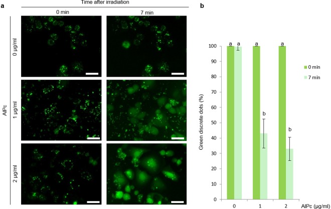
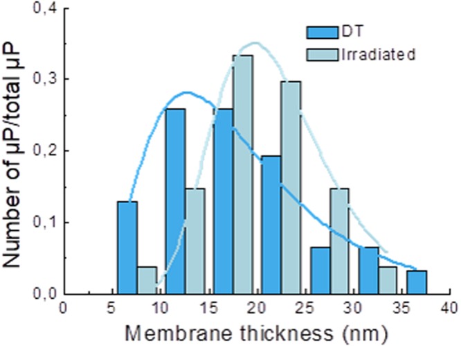
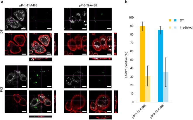
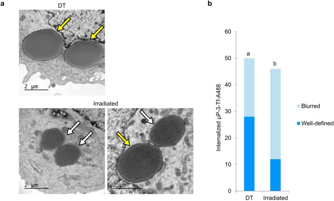
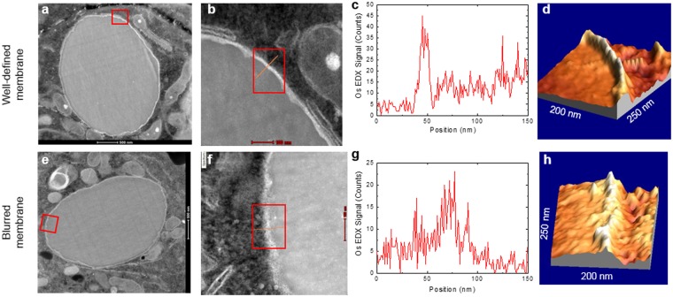
Therapeutic drug carriers can drive their cargo to their target cells. However, an obstacle is usually the entrapment of the drug inside the endolysosomal compartment, which physically impedes its actuation by the impossibility of reaching its molecular site of action. To overcome this hurdle, photochemical internalization (PCI) has been proposed, but the extent of PCI-induced membrane disruption and its capability to allow the release of microparticles is unknown. The aim of the present study was to determine if PCI allows the release of microparticles from the endolysosomal compartment to the cytosol and to analyze at the ultrastructural level the effect of PCI on the membrane surrounding the particles. Confocal microscope allowed us to detect that endolysosomal membranes suffered some disruption after PCI, evidenced by the diffusion of soluble transferrin from the endolysosomes to the cytosol and by a decrease of LAMP1-microparticles co-localization. Transmission electron microscopy (TEM) showed a decrease in the number of well-defined membranes around microparticles after PCI, and scanning TEM combined with energy dispersive x-ray revealed an increase in the width of endolysosomal membranes after treatment. These results suggest that endolysosomal membranes suffered an ultrastructure alteration after PCI, enough to liberate soluble transferrin but not the entire microparticles.
治疗性药物载体可以将其 cargo 输送到靶细胞。然而,通常的障碍是药物被内溶酶体隔室捕获,这通过使其无法到达其分子作用部位而物理上阻碍了其作用。为了克服这一障碍,已经提出了光化学内化(PCI),但 PCI 诱导的膜破坏的程度及其允许释放微粒的能力尚不清楚。本研究的目的是确定 PCI 是否允许微粒从内溶酶体隔室释放到细胞质中,并在超微结构水平上分析 PCI 对颗粒周围膜的影响。共聚焦显微镜使我们能够检测到 PCI 后内溶酶体膜受到了一些破坏,这可以通过可溶性转铁蛋白从内溶酶体扩散到细胞质以及 LAMP1-微粒的共定位减少来证明。透射电子显微镜 (TEM) 显示 PCI 后微粒周围的定义良好的膜数量减少,扫描 TEM 结合能量色散 X 射线显示处理后内溶酶体膜的宽度增加。这些结果表明,PCI 后内溶酶体膜发生了超微结构改变,足以释放可溶性转铁蛋白,但不能释放整个微粒。