Chen Ning, Su Wei, Cui Shu-Hui, Guo Jian, Duan Jia-Chuan, Li Hong-Xia, He Li
Department of Neurology, West China Hospital, Sichuan University, Chengdu, Sichuan Province, China.
Department of Neurology, West China Hospital; Department of Science & Technology, West China Hospital, Sichuan University, Chengdu, Sichuan Province, China.
Neural Regen Res. 2019 Jan;14(1):100-106. doi: 10.4103/1673-5374.243715.
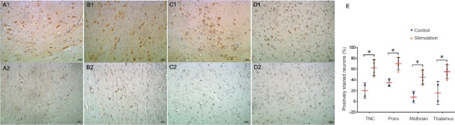
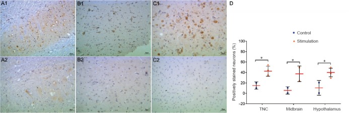
Several animal models of migraine have been established, and those based on trigeminovascular system activation are widely accepted. However, most of these models have been established on lower animals, such as rodents, and involve only a single administration of a noxious stimulus. In this study, an inflammatory soup (10 μL), consisting of prostaglandin E2 (0.2 mM), serotonin (2 mM), bradykinin (2 mM) and histamine (2 mM), was injected into the dura mater of conscious rhesus monkeys through an indwelling catheter. The infusion started on day 8 and was repeated every 3 days, for a total of six administrations, to induce neurogenic inflammation. We performed behavioral assessments and measured the expression of the oncogene c-fos, neuronal nitric oxide synthase (nNOS) and calcitonin gene related peptide (CGRP) in the trigeminal system and in multiple brain regions involved in pain processing by immunohistochemical staining. Compared with monkeys in the control group, three of the four animals in the inflammatory soup group displayed decreased motor behaviors, and two showed increased ipsilateral nose and mouth secretions during the stimulus period. Higher expression levels of c-fos, nNOS and CGRP were found in various brain areas of experimental animals compared with controls, including the trigeminal nucleus caudalis, thalamus, hypothalamus, midbrain, pons and other areas involved in pain perception. These results suggest that repeated inflammatory soup stimulation of the dura activates the trigeminovascular system and produces migraine-like pathological changes and abnormal behaviors in conscious rhesus monkeys.
已经建立了几种偏头痛动物模型,其中基于三叉神经血管系统激活的模型被广泛接受。然而,这些模型大多是在啮齿类等低等动物上建立的,并且仅涉及单次给予有害刺激。在本研究中,通过留置导管将由前列腺素E2(0.2 mM)、血清素(2 mM)、缓激肽(2 mM)和组胺(2 mM)组成的炎性液(10 μL)注入清醒恒河猴的硬脑膜。输注从第8天开始,每3天重复一次,共进行六次给药,以诱导神经源性炎症。我们进行了行为评估,并通过免疫组织化学染色测量了三叉神经系统以及参与疼痛处理的多个脑区中癌基因c-fos、神经元型一氧化氮合酶(nNOS)和降钙素基因相关肽(CGRP)的表达。与对照组的猴子相比,炎性液组的四只动物中有三只在刺激期间出现运动行为减少,两只出现同侧鼻口分泌物增加。与对照组相比,在实验动物的各个脑区,包括三叉神经尾核、丘脑、下丘脑、中脑、脑桥和其他参与疼痛感知的区域,发现c-fos、nNOS和CGRP的表达水平更高。这些结果表明,反复用炎性液刺激硬脑膜会激活三叉神经血管系统,并在清醒恒河猴中产生类似偏头痛的病理变化和异常行为。