Mustafi Sourajit M, Harezlak Jaroslaw, Kodiweera Chandana, Randolph Jennifer S, Ford James C, Wishart Heather A, Wu Yu-Chien
Department of Radiology and Imaging Sciences, Indiana University School of Medicine, Indianapolis, IN, USA.
Department of Epidemiology and Biostatistics, School of Public Health, Indiana University, Bloomington, IN, USA.
Neural Regen Res. 2019 Jan;14(1):114-123. doi: 10.4103/1673-5374.243716.
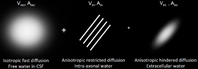
Multiple sclerosis is a neurodegenerative and inflammatory disease, a hallmark of which is demyelinating lesions in the white matter. We hypothesized that alterations in white matter microstructures can be non-invasively characterized by advanced diffusion magnetic resonance imaging. Seven diffusion metrics were extracted from hybrid diffusion imaging acquisitions via classic diffusion tensor imaging, neurite orientation dispersion and density imaging, and q-space imaging. We investigated the sensitivity of the diffusion metrics in 36 sets of regions of interest in the brain white matter of six female patients (age 52.8 ± 4.3 years) with multiple sclerosis. Each region of interest set included a conventional T2-defined lesion, a matched perilesion area, and normal-appearing white matter. Six patients with multiple sclerosis (n = 5) or clinically isolated syndrome (n = 1) at a mild to moderate disability level were recruited. The patients exhibited microstructural alterations from normal-appearing white matter transitioning to perilesion areas and lesions, consistent with decreased tissue restriction, decreased axonal density, and increased classic diffusion tensor imaging diffusivity. The findings suggest that diffusion compartment modeling and q-space analysis appeared to be sensitive for detecting subtle microstructural alterations between perilesion areas and normal-appearing white matter.
多发性硬化症是一种神经退行性和炎症性疾病,其特征是白质中的脱髓鞘病变。我们假设白质微结构的改变可以通过先进的扩散磁共振成像进行非侵入性表征。通过经典扩散张量成像、神经突方向分散和密度成像以及q空间成像,从混合扩散成像采集中提取了七个扩散指标。我们研究了六个患有多发性硬化症(年龄52.8±4.3岁)的女性患者脑白质中36组感兴趣区域的扩散指标的敏感性。每组感兴趣区域包括一个传统T2定义的病变、一个匹配的病变周围区域和外观正常的白质。招募了六名处于轻度至中度残疾水平的多发性硬化症患者(n = 5)或临床孤立综合征患者(n = 1)。患者表现出从外观正常的白质过渡到病变周围区域和病变的微结构改变,这与组织受限性降低、轴突密度降低以及经典扩散张量成像扩散率增加一致。研究结果表明,扩散隔室建模和q空间分析似乎对检测病变周围区域和外观正常的白质之间的细微微结构改变敏感。