Unit of Cardiology, Department of Medicine, Karolinska Institutet, Karolinska University Hospital, Stockholm 17176, Sweden.
Endocrinology and Diabetology, Department of Molecular Medicine and Surgery, Karolinska Institutet, Karolinska University Hospital, Stockholm 17176, Sweden.
Int J Mol Sci. 2018 Dec 7;19(12):3942. doi: 10.3390/ijms19123942.
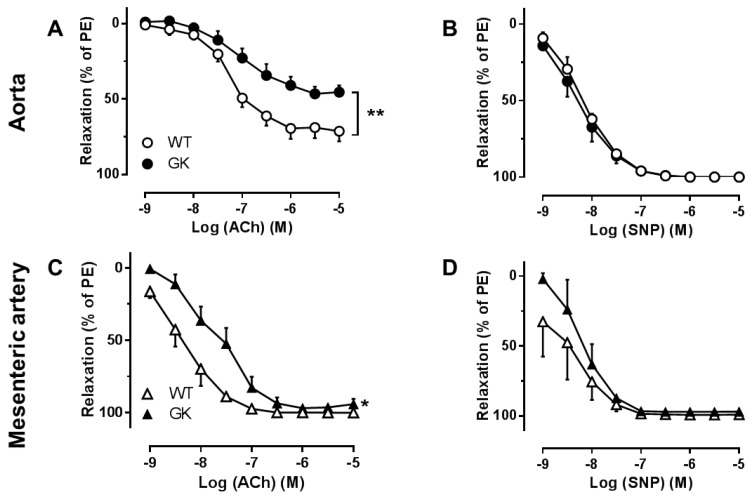
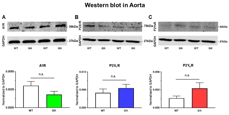
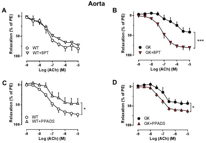
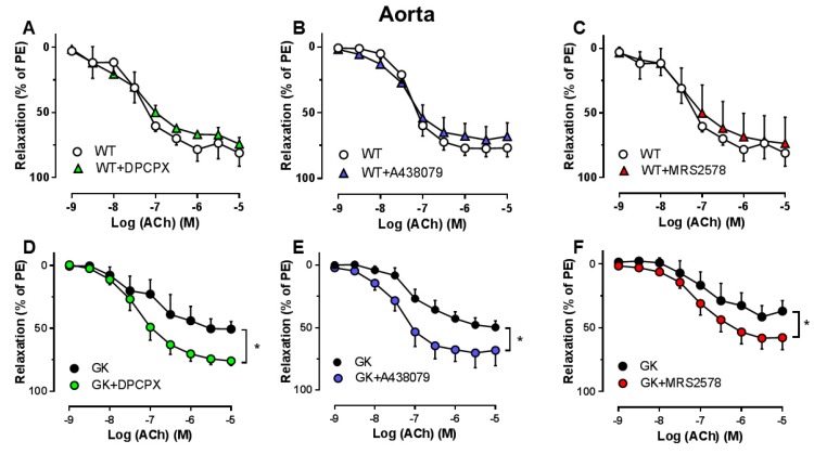
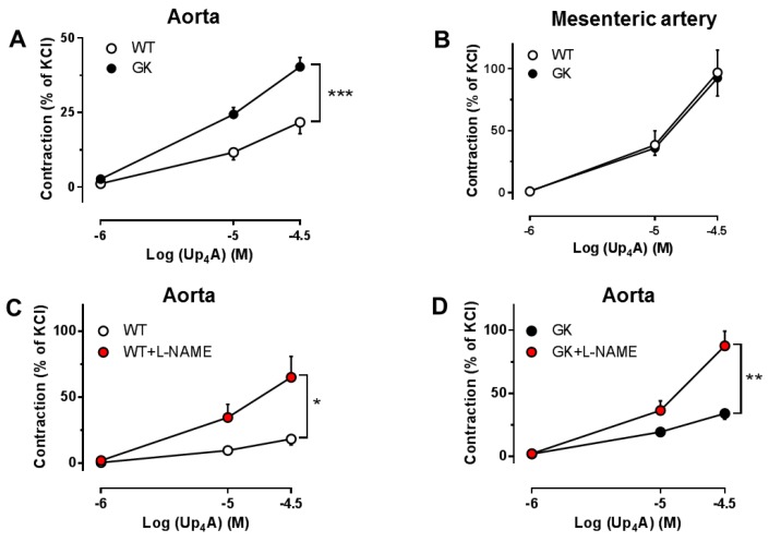
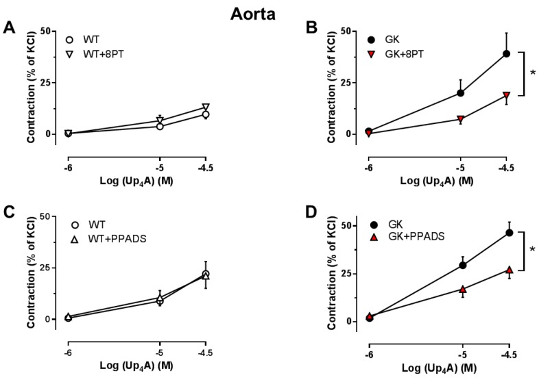
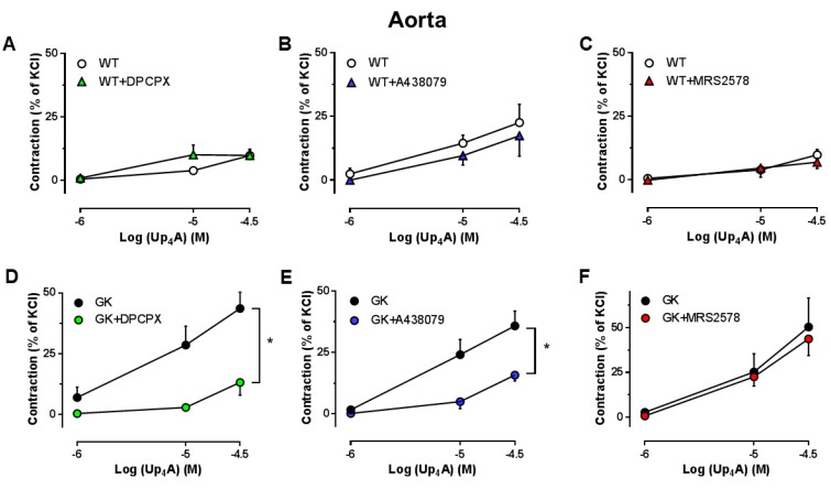
Purinergic signaling may be altered in diabetes accounting for endothelial dysfunction. Uridine adenosine tetraphosphate (Up₄A), a novel dinucleotide substance, regulates vascular function via both purinergic P1 and P2 receptors (PR). Up₄A enhances vascular contraction in isolated arteries of diabetic rats likely through P2R. However, the precise involvement of PRs in endothelial dysfunction and the vasoconstrictor response to Up₄A in diabetes has not been fully elucidated. We tested whether inhibition of PRs improved endothelial function and attenuated Up₄A-mediated vascular contraction using both aortas and mesenteric arteries of type 2 diabetic (T2D) Goto Kakizaki (GK) rats vs. control Wistar (WT) rats. Endothelium-dependent (EDR) but not endothelium-independent relaxation was significantly impaired in both aortas and mesenteric arteries from GK vs. WT rats. Non-selective inhibition of P1R or P2R significantly improved EDR in aortas but not mesenteric arteries from GK rats. Inhibition of A1R, P2X₇R, or P2Y₆R significantly improved EDR in aortas. Vasoconstrictor response to Up₄A was enhanced in aortas but not mesenteric arteries of GK vs. WT rats via involvement of A1R and P2X₇R but not P2Y₆R. Depletion of major endothelial component nitric oxide enhanced Up₄A-induced aortic contraction to a similar extent between WT and GK rats. No significant differences in protein levels of A1R, P2X₇R, and P2Y₆R in aortas from GK and WT rats were observed. These data suggest that altered PR sensitivity accounts for endothelial dysfunction in aortas in diabetes. Modulating PRs may represent a potential therapy for improving endothelial function.
嘌呤能信号可能在糖尿病中发生改变,从而导致内皮功能障碍。尿苷腺苷四磷酸 (Up₄A) 是一种新型二核苷酸物质,通过嘌呤能 P1 和 P2 受体 (PR) 调节血管功能。Up₄A 通过 P2R 增强糖尿病大鼠离体动脉的血管收缩。然而,PR 在糖尿病内皮功能障碍和 Up₄A 介导的血管收缩中的确切作用尚未完全阐明。我们使用 2 型糖尿病 (T2D) Goto Kakizaki (GK) 大鼠与对照 Wistar (WT) 大鼠的主动脉和肠系膜动脉来测试 PR 抑制是否改善内皮功能并减弱 Up₄A 介导的血管收缩。与 WT 大鼠相比,GK 大鼠的主动脉和肠系膜动脉中内皮依赖性舒张 (EDR) 但非内皮非依赖性舒张明显受损。非选择性 P1R 或 P2R 抑制显著改善了 GK 大鼠主动脉的 EDR,但对肠系膜动脉无影响。A1R、P2X₇R 或 P2Y₆R 的抑制显著改善了 GK 大鼠主动脉的 EDR。通过 A1R 和 P2X₇R 的参与,但不通过 P2Y₆R,Up₅A 对 GK 大鼠主动脉的血管收缩反应增强,但对 WT 大鼠肠系膜动脉无影响。主要内皮成分一氧化氮的耗竭增强了 WT 和 GK 大鼠之间 Up₅A 诱导的主动脉收缩。在 GK 和 WT 大鼠的主动脉中,A1R、P2X₇R 和 P2Y₆R 的蛋白水平没有明显差异。这些数据表明,PR 敏感性的改变导致了糖尿病中主动脉的内皮功能障碍。调节 PR 可能是改善内皮功能的一种潜在治疗方法。