Department of Surgery, Jichi Medical University, 3311-1 Yakushiji, Shimotsuke, Tochigi, Japan.
Department of Diagnostic Pathology, Jichi Medical University, 3311-1 Yakushiji, Shimotsuke, Tochigi, Japan.
BMC Cancer. 2018 Dec 13;18(1):1249. doi: 10.1186/s12885-018-5165-0.
Thymomas are typically slow-growing tumors and AB type thymomas are considered no/low risk tumors with a better prognosis. Extra-thoracic metastases are extremely rare. To the best of our knowledge, no patient with an isolated splenic metastasis from a thymoma has been reported. We report a patient who underwent laparoscopic splenectomy for a slow-growing, isolated splenic metastasis, eight years after thymectomy.
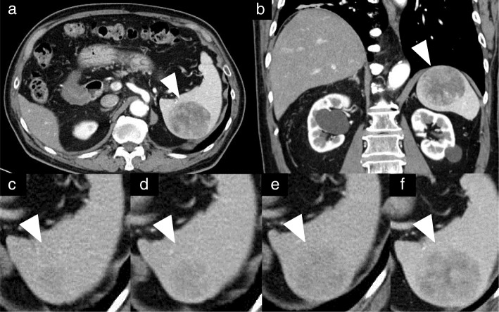
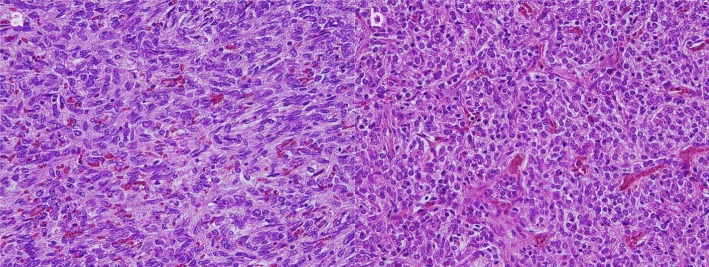
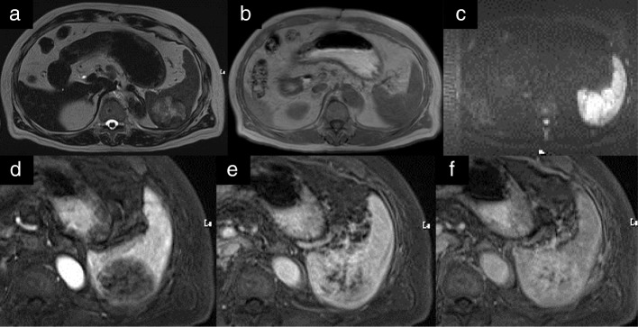
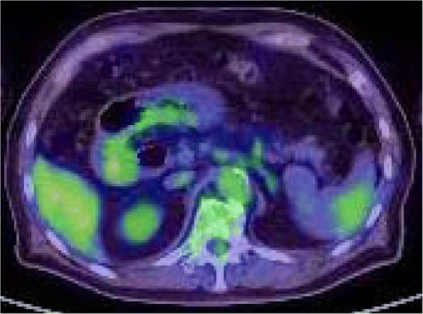
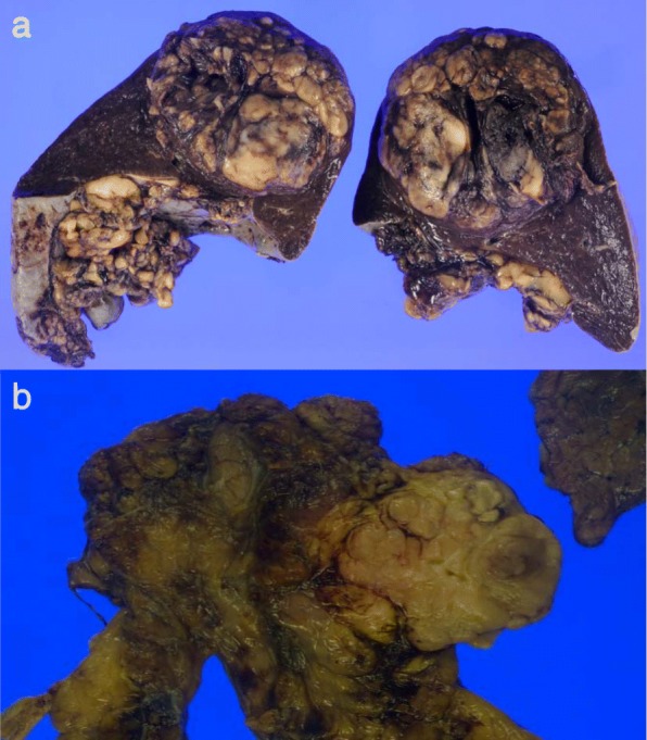
The patient is a 78-year-old man. Eight years previously, the patient underwent extended thymectomy and postoperative radiation therapy for a thymoma. Five years after thymectomy, a nodule appeared in the spleen, and the lesion enlarged gradually for three years thereafter. The patient was referred for further examination and treatment. Computed tomography scan showed a sharply circumscribed 50 mm tumor slightly hypodense and heterogeneous lesion in the spleen. On T2-weighted images on Magnetic Resonance Imaging, the tumor had high intensity, equivalent to or slightly lower than that on T1-weighted images, and no decrease on diffusion-weighted images. The tumor was multinodular and showed a low-signal spoke-wheel sign in the margin, enhanced gradually in the dynamic study. Positron emission tomography-CT scan, showed relatively low accumulation. Surgical resection was undertaken, and pathological examination showed metastatic thymoma. The patient is without recurrence and has no other symptoms three years after splenectomy.
This is the first report of an isolated splenic metastasis from a thymoma. Further cases are needed to standardize this surgery for such lesions.
胸腺瘤通常生长缓慢,AB 型胸腺瘤被认为是低风险肿瘤,预后较好。胸外转移极为罕见。据我们所知,尚无胸腺瘤孤立性脾转移的报道。我们报告一例患者,在胸腺切除术后 8 年,因生长缓慢、孤立性脾转移而行腹腔镜脾切除术。
患者为 78 岁男性。8 年前,患者因胸腺瘤行扩大胸腺切除术和术后放疗。胸腺切除术后 5 年,脾内出现结节,此后 3 年内病变逐渐增大。患者因进一步检查和治疗而被转介。计算机断层扫描显示脾脏内有一个边界清楚的 50mm 肿瘤,呈轻度低密和不均匀病变。磁共振成像上 T2 加权图像显示肿瘤呈高信号,与 T1 加权图像等或稍低,弥散加权图像上无下降。肿瘤呈多结节状,边缘呈低信号辐轮征,在动态研究中逐渐增强。正电子发射断层扫描 - 计算机断层扫描显示相对低积聚。行手术切除,病理检查显示为转移性胸腺瘤。患者在脾切除术后 3 年无复发且无其他症状。
这是首例胸腺瘤孤立性脾转移的报道。需要进一步的病例来规范此类病变的手术治疗。