Department of Diagnostic Medicine, Dell Medical School, University of Texas at Austin, Austin, TX
Division of Diabetes, Endocrinology, and Metabolism, Department of Medicine, Vanderbilt University Medical Center, Nashville, TN.
Diabetes Care. 2019 Feb;42(2):248-257. doi: 10.2337/dc18-1507. Epub 2018 Dec 14.
This study investigated the temporal dynamics of pancreas volume and microstructure in children and adolescents with recent-onset type 1 diabetes (T1D) and individuals without diabetes, including a subset expressing autoantibodies associated with the early stages of T1D.
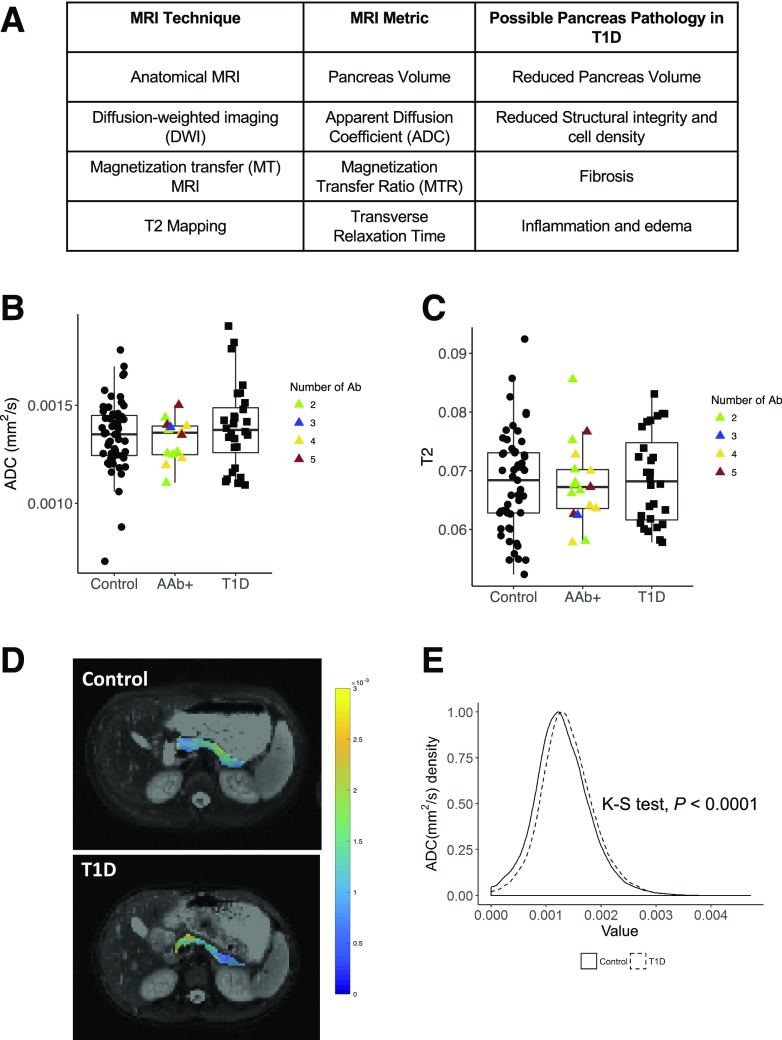
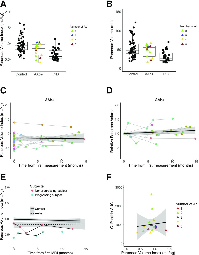
MRI was performed in individuals with recent-onset stage 3 T1D ( = 51; median age 13 years) within 100 days after diagnosis (mean 67 days), 6 months, and 1 year postdiagnosis. Longitudinal MRI measurements were also made in similarly aged control participants ( = 57) and in autoantibody-positive individuals without diabetes ( = 20). The MRI protocol consisted of anatomical imaging to determine pancreas volume and quantitative MRI protocols interrogating tissue microstructure and composition.
Within 100 days of diabetes onset, individuals with T1D had a smaller pancreas (median volume 28.6 mL) than control participants (median volume 48.4 mL; < 0.001), including when normalized by individual weight ( < 0.001). Longitudinal measurements of pancreas volume increased in control participants over the year, consistent with adolescent growth, but pancreas volume declined over the first year after T1D diagnosis ( < 0.001). In multiple autoantibody-positive individuals, the pancreas volume was significantly larger than that of the T1D cohort ( = 0.017) but smaller than that of the control cohort ( = 0.04). Diffusion-weighted MRI showed that individuals with recent-onset T1D had a higher apparent diffusion coefficient ( = 0.012), suggesting a loss of cellular structural integrity, with heterogeneous pancreatic distribution.
These results indicate that pancreas volume is decreased in stages 1, 2, and 3 of T1D and decreases during the first year after diabetes onset and that this loss of pancreatic volume is accompanied by microstructural changes.
本研究旨在探讨近期发病 1 型糖尿病(T1D)患儿和非糖尿病个体胰腺体积和微观结构的时间动态变化,其中包括一组表达与 T1D 早期阶段相关的自身抗体的个体。
在诊断后 100 天内(平均 67 天)、6 个月和 1 年时,对近期发病 3 期 T1D 患者(n=51;中位年龄 13 岁)进行 MRI 检查。还对年龄匹配的对照组参与者(n=57)和无糖尿病的自身抗体阳性个体(n=20)进行了纵向 MRI 测量。MRI 方案包括解剖成像以确定胰腺体积和定量 MRI 方案以检测组织微观结构和组成。
在糖尿病发病后 100 天内,T1D 患者的胰腺体积(中位数 28.6mL)明显小于对照组(中位数 48.4mL;<0.001),包括按个体体重校正后(<0.001)。在对照组参与者中,胰腺体积在一年中会随着青少年生长而增加,但在 T1D 诊断后的第一年,胰腺体积会下降(<0.001)。在多个近期发病的自身抗体阳性个体中,胰腺体积明显大于 T1D 队列(=0.017),但小于对照组(=0.04)。弥散加权 MRI 显示,近期发病的 T1D 患者的表观扩散系数较高(=0.012),这表明细胞结构完整性丧失,胰腺分布不均匀。
这些结果表明,T1D 的 1 期、2 期和 3 期胰腺体积减少,并且在糖尿病发病后的第一年减少,而这种胰腺体积的损失伴随着微观结构的变化。