Department of Chemistry and Biochemistry, Kent State University at Trumbull, Warren, Ohio.
Princeton Neuroscience Institute, Princeton University, Princeton, New Jersey.
Brain Behav. 2018 Dec;8(12):e01165. doi: 10.1002/brb3.1165. Epub 2018 Nov 22.
We compared the integrity of white matter (WM) microstructure to the course of recovery in athletes who sustained one sports-related concussion (SRC), assessing individual longitudinal changes in WM fiber tracts following SRC using pre- and post-injury measurements.
Baseline diffusion tensor imaging (DTI) scans and neuropsychological tests were collected on 53 varsity contact-sport college athletes. Participants (n = 13) who subsequently sustained an SRC underwent DTI scans and neuropsychological testing at 2 days, 2 weeks, and 2 months following injury.
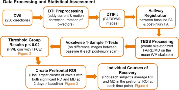
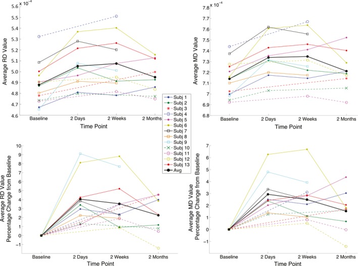
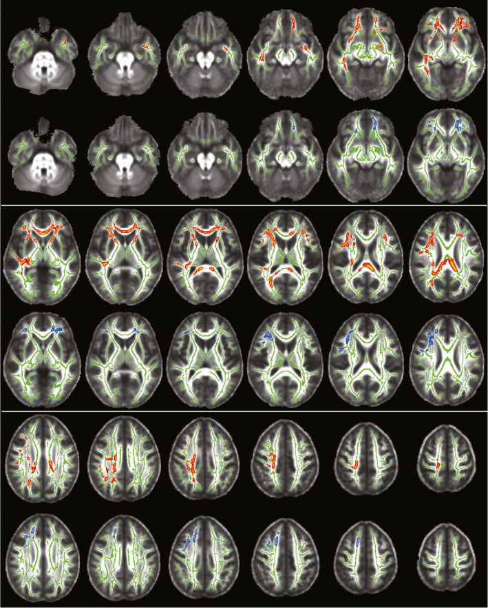
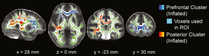
Relying on tract-based spatial statistics (TBSS) analyses, we found that radial diffusivity (RD) and mean diffusivity (MD) were significantly increased at 2 days post-injury compared to the same-subject baseline (corrected p < 0.02). These alterations were visible in anterior/posterior WM regions spanning both hemispheres, demonstrating a diffuse pattern of injury after concussion. Implicated WM fiber tracts at 2 days include the following: right superior/inferior longitudinal fasciculus; right/left inferior fronto-occipital fasciculus; right corticospinal tract; right acoustic radiation; right/left anterior thalamic radiations; right/left uncinate fasciculus; and forceps major/minor. At 2 weeks post-injury, persistently elevated RD and MD were observed solely in prefrontal portions of WM fiber tracts (using same-subject contrasts). No significant differences were found for FA in any of the post-injury comparisons to baseline. Plots of individual subject RD and MD in prefrontal WM demonstrated homogenous increases from baseline to just after SRC; thereafter, trajectories became more variable. Most subjects' diffusivity values remained elevated at 2 months post-injury relative to their own baseline. Over the 2-month period after SRC, recovery of WM fiber tracts appeared to follow a posterior-to-anterior trend, paralleling the posterior-anterior pattern of WM maturation previously identified in the normal population.
These results suggest greater vulnerability of prefrontal regions to SRC, underline the importance of an individualized approach to concussion management, and show promise for using RD and MD for imaging-based diagnosis of SRC.
我们比较了脑白质(WM)微观结构的完整性与运动员遭受单一运动相关脑震荡(SRC)后的恢复过程,通过 SRC 前后的测量评估 WM 纤维束在 SRC 后的个体纵向变化。
对 53 名大学生接触性运动项目运动员进行了基线扩散张量成像(DTI)扫描和神经心理学测试。随后发生 SRC 的参与者(n=13)在受伤后 2 天、2 周和 2 个月接受了 DTI 扫描和神经心理学测试。
基于基于纤维束的空间统计学(TBSS)分析,我们发现与同一受试者基线相比,损伤后 2 天的各向异性分数(FA)值降低,而径向扩散系数(RD)和平均扩散系数(MD)显著升高(校正后 p<0.02)。这些改变在跨越两个半球的前后 WM 区域可见,表明脑震荡后存在弥漫性损伤模式。在 2 天内损伤的 WM 纤维束包括:右侧上下纵束;右侧/左侧下额枕束;右侧皮质脊髓束;右侧听辐射;右侧/左侧前丘脑辐射;右侧/左侧钩束;以及主要/次要内囊。在受伤后 2 周时,仅在前额叶 WM 纤维束的部位观察到持续升高的 RD 和 MD(使用同一受试者对比)。在与基线的任何损伤后比较中,FA 均无显著差异。在前额叶 WM 中个体受试者 RD 和 MD 的变化图显示从基线到 SRC 后立即呈均匀增加;此后,轨迹变得更加多变。大多数受试者的弥散值在 SRC 后 2 个月仍高于自身基线。在 SRC 后 2 个月期间,WM 纤维束的恢复似乎遵循从后向前的趋势,与先前在正常人群中确定的 WM 成熟的后向前模式平行。
这些结果表明,前额区域对 SRC 更敏感,强调了对脑震荡管理采用个体化方法的重要性,并为使用 RD 和 MD 进行基于成像的 SRC 诊断提供了希望。