Department of Molecular Imaging and Theranostics, National Institute of Radiological Sciences, National Institutes for Quantum and Radiological Science and Technology (QST), Chiba, Japan.
Group of Quantum-State Controlled MRI, QST, Chiba, Japan.
Front Neural Circuits. 2018 Dec 6;12:110. doi: 10.3389/fncir.2018.00110. eCollection 2018.
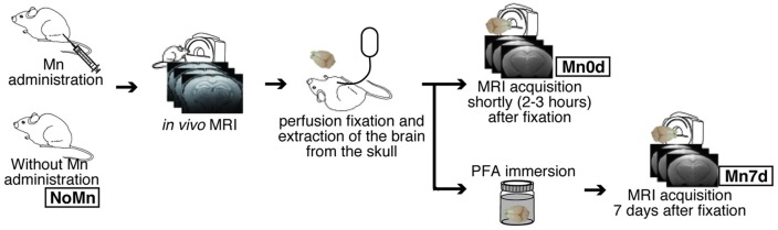
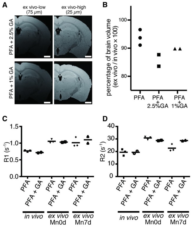
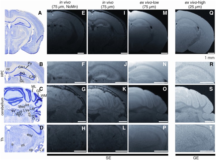
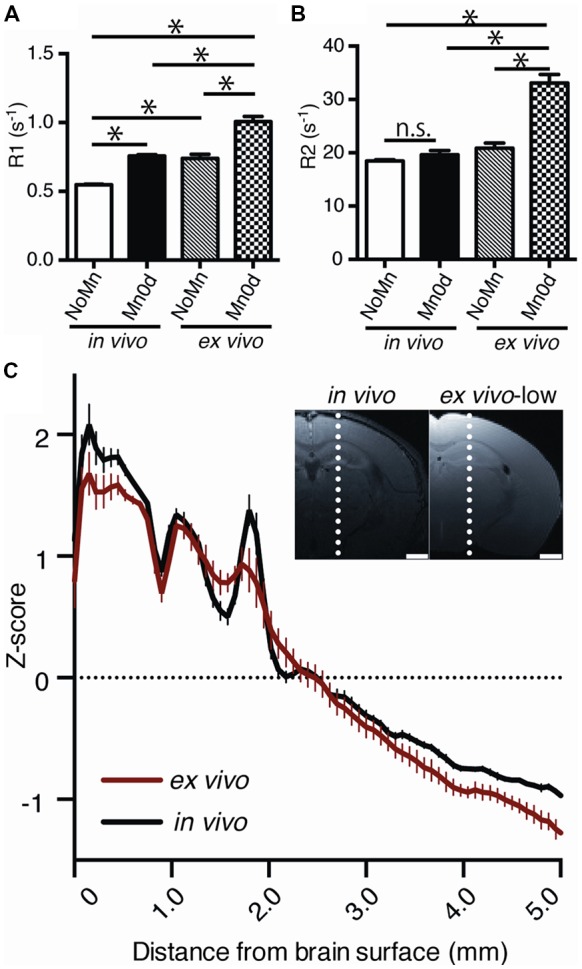
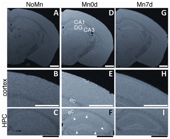
MRI observations following administration of Mn [manganese (Mn)-enhanced MRI, MEMRI] have been used as an excellent morphological and functional MRI tool for preclinical studies. To detect brain three-dimensional (3D) microstructures, we improved the MEMRI method for mouse brains after Mn administration and obtained high-resolution MRIs using a cryogenic radiofrequency (RF) coil. Male C57BL/6 mice ( = 8) were injected with 50 mM MnCl intravenously and MEMRIs of the brain were acquired after 24 h, followed by perfusion fixation with a 4% paraformaldehyde (PFA) solution. High-resolution 25-μm isotropic MRIs were successfully acquired from the extracted brain tissue and could identify the brain microstructures, especially in the hippocampus [the pyramidal cell layer through CA1-3 and the dentate gyrus (DG) granular layers (GLs)], cell layers of cerebellum, three sub-regions of the deep cerebellar nucleus, and white matter (WM) structures [e.g., the fasciculus retroflexus (fr) and optic tract in the thalamus]. The following technical conditions were also examined: (i) the longitudinal stability of Mn-enhanced tissue after administration; and (ii) the effects of mixing glutaraldehyde (GA) with the fixative solution for the preservation of MEMRI contrast. Our results indicate that MEMRI observations made shortly after fixation maintain the contrast observed . This research will be useful for non-destructive whole-brain pathological analysis.
Mn[锰(Mn)增强磁共振成像,MEMRI]给药后的 MRI 观察已被用作临床前研究的出色形态和功能 MRI 工具。为了检测大脑三维(3D)微观结构,我们改进了 Mn 给药后小鼠大脑的 MEMRI 方法,并使用低温射频(RF)线圈获得了高分辨率 MRI。雄性 C57BL/6 小鼠(n=8)静脉注射 50 mM MnCl2,24 小时后进行脑 MEMRI,并随后用 4%多聚甲醛(PFA)溶液进行灌注固定。从提取的脑组织中成功获得了高分辨率 25-μm 各向同性 MRI,可以识别大脑微观结构,特别是在海马体[CA1-3 锥体细胞层和齿状回(DG)颗粒层]、小脑细胞层、小脑深部核的三个亚区和白质(WM)结构[例如,丘脑中的折返束(fr)和视束]。还检查了以下技术条件:(i)给药后 Mn 增强组织的纵向稳定性;和(ii)戊二醛(GA)与固定液混合对保存 MEMRI 对比的影响。我们的结果表明,固定后不久进行的 MEMRI 观察保持了观察到的对比度。这项研究对于无损全脑病理分析将非常有用。