Molecular Biophysics Laboratory, Photonics Center and Department of Physics, Boston University, Boston, Massachusetts, United States of America.
Department of Biophysical Organic Chemistry, Leiden Institute of Chemistry, Leiden UniversityAR Leiden, The Netherlands.
PLoS One. 2018 Dec 26;13(12):e0209506. doi: 10.1371/journal.pone.0209506. eCollection 2018.
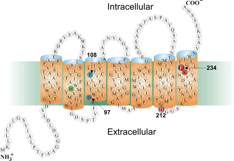
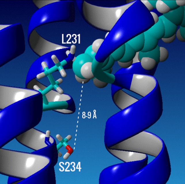
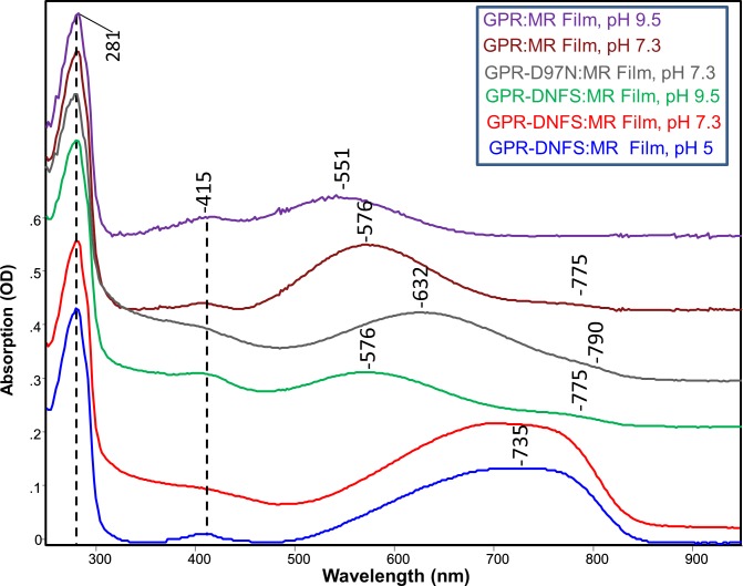
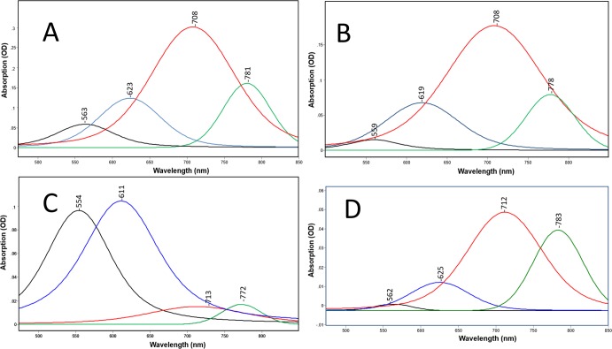
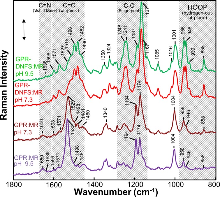
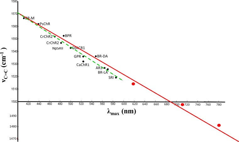
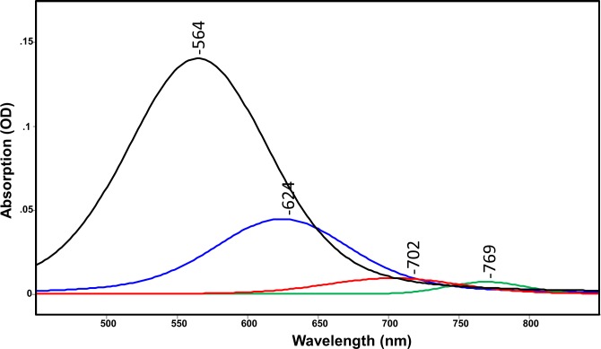
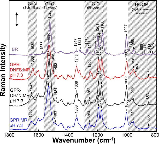
Microbial rhodopsins have become an important tool in the field of optogenetics. However, effective in vivo optogenetics is in many cases severely limited due to the strong absorption and scattering of visible light by biological tissues. Recently, a combination of opsin site-directed mutagenesis and analog retinal substitution has produced variants of proteorhodopsin which absorb maximally in the near-infrared (NIR). In this study, UV-Visible-NIR absorption and resonance Raman spectroscopy were used to study the double mutant, D212N/F234S, of green absorbing proteorhodopsin (GPR) regenerated with MMAR, a retinal analog containing a methylamino modified β-ionone ring. Four distinct subcomponent absorption bands with peak maxima near 560, 620, 710 and 780 nm are detected with the NIR bands dominant at pH <7.3, and the visible bands dominant at pH 9.5. FT-Raman using 1064-nm excitation reveal two strong ethylenic bands at 1482 and 1498 cm-1 corresponding to the NIR subcomponent absorption bands based on an extended linear correlation between λmax and γC = C. This spectrum exhibits two intense bands in the fingerprint and HOOP mode regions that are highly characteristic of the O640 photointermediate from the light-adapted bacteriorhodopsin photocycle. In contrast, 532-nm excitation enhances the 560-nm component, which exhibits bands very similar to light-adapted bacteriorhodopsin and/or the acid-purple form of bacteriorhodopsin. Native GPR and its mutant D97N when regenerated with MMAR also exhibit similar absorption and Raman bands but with weaker contributions from the NIR absorbing components. Based on these results it is proposed that the NIR absorption in GPR-D212N/F234S with MMAR arises from an O-like chromophore, where the Schiff base counterion D97 is protonated and the MMAR adopts an all-trans configuration with a non-planar geometry due to twists in the conjugated polyene segment. This configuration is characterized by extensive charge delocalization, most likely involving nitrogens atoms in the MMAR chromophore.
微生物视紫红质已成为光遗传学领域的重要工具。然而,由于可见光在生物组织中的强吸收和散射,有效的活体光遗传学在许多情况下受到严重限制。最近,通过视蛋白定点突变和类似视黄醛取代的组合,产生了吸收最大值在近红外(NIR)的变构菌视紫红质(proteorhodopsin)变体。在这项研究中,使用 UV-可见-NIR 吸收和共振拉曼光谱研究了与 MMAR 再生的绿色吸收变构菌视紫红质(GPR)的双突变体 D212N/F234S,MMAR 是一种含有甲氨基修饰的β-紫罗兰酮环的视黄醛类似物。在 pH <7.3 时,检测到四个明显的亚组分吸收带,峰值约为 560、620、710 和 780nm,NIR 带占主导地位;在 pH 9.5 时,可见带占主导地位。使用 1064nm 激发的 FT-Raman 显示两个强的乙基烯带在 1482 和 1498cm-1,对应于 NIR 亚组分吸收带,基于 λmax 和 γC = C 之间的扩展线性相关性。该光谱在指纹和 HOOP 模式区域显示两个强烈的带,高度特征于从光适应菌视紫红质光循环的 O640 光中间体。相比之下,532nm 激发增强了 560nm 成分,其显示出与光适应菌视紫红质和/或菌视紫红质的酸紫色形式非常相似的带。用 MMAR 再生的天然 GPR 及其突变体 D97N 也表现出类似的吸收和拉曼带,但 NIR 吸收成分的贡献较弱。基于这些结果,提出 GPR-D212N/F234S 与 MMAR 的 NIR 吸收来自于 O 样生色团,其中席夫碱反离子 D97 质子化,并且 MMAR 由于共轭多烯段的扭曲而采用全反式构型和非平面几何形状。这种构象的特征是广泛的电荷离域,很可能涉及 MMAR 生色团中的氮原子。